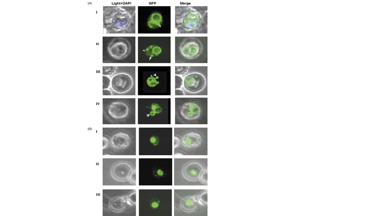
Trafficking of falcipain 2-GFP fusion proteins. Confocal microscope images showing expression and trafficking of Fal2-GFP fusion proteins at trophozoite
stage of Fal2-S1 (A) and Fal2-S2 (B) transgenic P. falciparum parasites. The first image in each set (left to right) represents the bright field image, the second is the fluorescence signal from the GFP chimeric protein and an overlay of these two images is shown in the third image. In both the parasite lines, the chimeric GFP is visible in parasitophorous vacuole (shown with arrow heads), and in the extension or evaginations of the parasitophorous vacuole (shown with pointed arrows). Since falcipain-2 has been shown to be a principal trophozoite stage cysteine protease, we studied in detail the distribution of chimeric proteins at trophozoite stage of P. falciparum (36 h post invasion). In Fal2-S1 transgenic line, the GFP fluorescence was detected in ER around the nucleus, (A, panel II and panel IV), in food vacuole membrane (A, panel I, II, III and IV), in parasitophorous vacuole and in protrusions in the erythrocyte cytoplasm, which apparently arises from PVM (A, panel I, III and IV). These protrusions appear to be part of tubovesicular network that lies in erythrocyte cytoplasm. The Fal2-S2 transgenic line showed distribution of GFP in ER, within food vacuole and in small vesicles/foci as seen in Fal2-S1 construct (B). Some of these foci seem to be originating from parasitophorous vacuole, while others seem to be fusing with the food vacuole (A, panel II and III; B, panel II and III.
Dasaradhi PV, Mohmmed A, Kumar A, Hossain MJ, Bhatnagar RK, Chauhan VS, Malhotra P. A role of falcipain-2, principal cysteine proteases of Plasmodium falciparum in merozoite egression. Biochem Biophys Res Commun. 2005 336(4):1062-8.
Other associated proteins
| PFID | Formal Annotation |
|---|---|
| PF3D7_1115700 | cysteine proteinase falcipain 2a falcipain 2 |