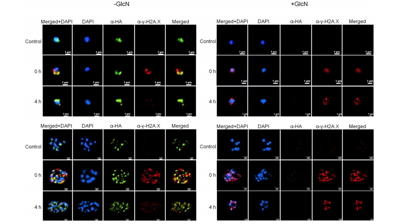
Dynamics of PfH2A phosphorylation in P. falciparum nucleus following X-ray induced DNA damage in the presence or absence of PfSR1 expression. Immuno-fluorescence imaging of γ-PfH2A (red) and PfSR1 (green) in the nucleus of early (upper panels) and late stages (lower panels) PfSR1-glmS parasites grown 72h either on regular media (-GlcN, left panels) or media supplemented with 5mM GlcN (+ GlcN, right panels) to knockdown PfSR1 expression. Parasite were exposed to X-ray irradiation (3000 rad) and the association between PfSR1 and the
γ-PfH2A foci formation was imaged before irradiation (Control), 15 minutes after exposure (0h), and 4 hour post (4h) X-ray irradiation. DNA is stained with DAPI, scale bar 1 µm. in parasites in which PfSR1 expression is knocked-down, PfRad51 does not accumulate in the nucleus and thus the levels of γ-PfH2A does not decrease over time.
Goyal M, Singh BK, Simantov K, Kaufman Y, Eshar S, Ron D. An SR protein is essential for activating DNA repair in malaria parasites. J Cell Sci. 2021 jcs.258572.
Other associated proteins
| PFID | Formal Annotation |
|---|---|
| PF3D7_1131100 | serpentine receptor 1, putative, PfSr1 |