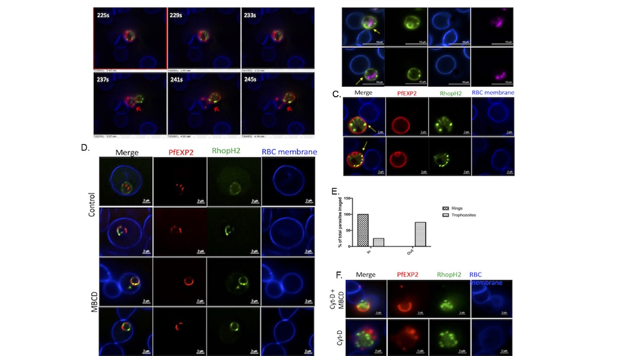
Trophozoites extrude out from erythrocyte upon treatment with MbCD. (A) Still frames from Video S1 in the supplemental material from time-lapse video microscopy. Parasites extrude out from the erythrocyte with the PVM still tethered to the erythrocyte membrane without causing the lysis of erythrocytes (indicated by arrows). (B) Representative images (.50) of the PfVP1-mNG line after MbCD treatment. Nucleus is stained with SYTO deep red (pink). (C) Representative images (.50) of the NF54 RhopH2/Exp2 line after MbCD treatment. Erythrocyte membrane is stained with WGA-Alexa 350 (blue). Infected erythrocytes were subjected to live fluorescence microscopy after treatment with MbCD. Parasites extrude out from the erythrocyte without causing lysis of the erythrocyte with their PPM (B) and PVM (C) still attached to the erythrocyte as indicated by the arrows. (D) Representative images of erythrocytes infected with ring stages of the NF54 RhopH2/Exp2 line treated with MbCD. Ring-stage parasites do not extrude out from erythrocytes. (E) Quantification of parasites remaining inside/outside erythrocytes after MbCD treatment was carried out by examining at least 100 different images of individual parasites for each experimental condition (100 for ring stages and 330 for trophozoites). (F, Top) Treatment of NF54 RhopH2/Exp2 trophozoites with 0.5 mM Cyt-D for 45 min prior to MbCD treatment does not inhibit parasite extrusion. (F, Bottom) Treatment with 0.5 mM Cyt-D alone does not cause extrusion of trophozoites but did alter PVM morphology (as indicated by arrows). (G, Left) Treatment with MbCD causes extrusion of late-stage P. falciparum- without causing erythrocyte rupture, unlike the control group where the trophozoite is still inside the erythrocyte (scale bar, 2 mM; direct magnification, x5,000). (G, Right) Zoomed-in image of parasite extruded out of erythrocyte after MbCD treatment shows the PVM ruptured at multiple places indicated by black arrows. Electron microscopy of extruded P. falciparum. (G, Left) Lower-magnification views of control and MbCD-treated trophozoites. A higher-magnification view of an extruded parasite (right) shows that the PVM is compromised at multiple places (arrows) (scale bar, 500 nM; direct magnification, x12,000).
Ahiya AI, Bhatnagar S, Morrisey JM, Beck JR, Vaidya AB. Dramatic Consequences of Reducing Erythrocyte Membrane Cholesterol on Plasmodium falciparum. Microbiol Spectr. 2022; 10(1 ):e0015822.
Other associated proteins
| PFID | Formal Annotation |
|---|---|
| PF3D7_0929400 | high molecular weight rhoptry protein 2 |