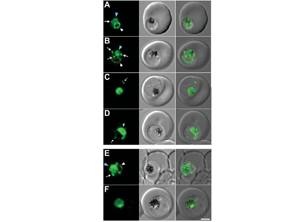
(A–D) 3D7 and (E, F) Dd2 transfectants expressing PM2–GFP were imaged at the young to mid trophozoite (A, B, E) or mature trophozoite to schizont (C, D, F) stage. The images (left to right) represent a GFP fluorescence image, a DIC (differential interference contrast) image and an overlay of these images. DV-located GFP is indicated with a blue arrowhead. ER-located GFP is indicated with white arrowheads, while cytostomal vesicles are indicated with white arrows. Scale bar, 2 mm
Klonis N, Tan O, Jackson K, Goldberg D, Klemba M, Tilley L. Evaluation of pH during cytostomal endocytosis and vacuolar catabolism of haemoglobin in Plasmodium falciparum. Biochem J. 2007 407(3):343-54.