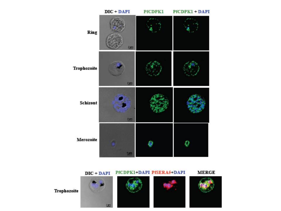
PfCDPK1 is expressed throughout erythrocytic schizogony as detected by by immunofluorescence assay (IFA). PfCDPK1 was detected in blood stages of P. falciparum by IFA using anti-PfCDPK1 mouse sera and Alexa Fluor 488-conjugated anti-mouse IgG goat sera (green). Anti-SERA5 rabbit sera and Alexa Fluor 594-conjugated anti-rabbit IgG goat sera (red) were used to localize the parasitophorous vacuole (PV) protein SERA5. Nuclear DNA was counterstained with DAPI (blue). Bright field (DIC) merged with DAPI (DIC + DAPI), fluorescence images detecting PfCDPK1 (PfCDPK1) and merged fluorescence images detecting PfCDPK1 and DAPI, PfSERA5 and DAPI or PfCDPK1, PfSERA5 and DAPI (Merge) are shown. Bar represents 2 mm (ring, trophozoite, schizont) or 1 μm (merozoite). PfCDPK1 localized to the rim of the infected erythrocyte and appeared to be associated with the erythrocyte membrane in rings and trophozoites (lower panel). In addition, PfCDPK1 was found in the parasitophorous vacuole (PV) and co-localized with SERA5, a known PV protein in trophozoites. Inmerozoites, PfCDPK1 was primarily found at the boundary of the merozoite at the merozoite plasma membrane
Bansal A, Singh S, More KR, Hans D, Nangalia K, Yogavel M, Sharma A, Chitnis CE. Characterization of Plasmodium falciparum calcium dependent protein kinase 1 (PfCDPK1) and its role in microneme secretion during erythrocyte invasion. J Biol Chem. 2013 288(3):1590-602.
Other associated proteins
| PFID | Formal Annotation |
|---|---|
| PF3D7_0207600 | serine repeat antigen 5 |