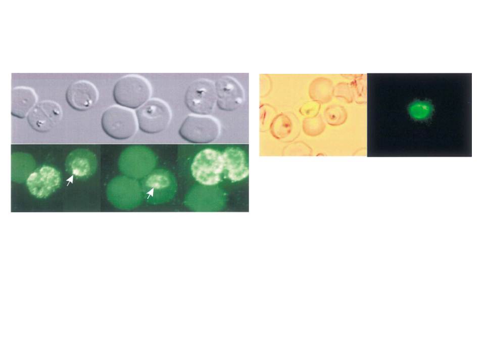
Left: Immunofluorescence microscopy of erythrocytes infected with trophozoite stage P. falciparum. Bright fields showing parasite morphology were collected by differential interference contrast. Fluorescence images were taken after reaction with antibody PABHK and FITC-conjugated secondary antibody. Arrows indicate an intense spot of fluorescence frequently observed. PfVP1 is localized to the parasite plasma membrane
Right: Epifluorescence of P. falciparum transfected with GFP gene fusions. Trophozoite transfected with PfVP1–GFP gene fusion showing a membrane bound parasite surface distribution and a bright spot of fluorescence.
McIntosh MT, Drozdowicz YM, Laroiya K, Rea PA, Vaidya AB. Two classes of plant-like vacuolar-type H(+)-pyrophosphatases in malaria parasites. Mol Biochem Parasitol. 2001 114:183-95. Copyright Elsevier 2009.