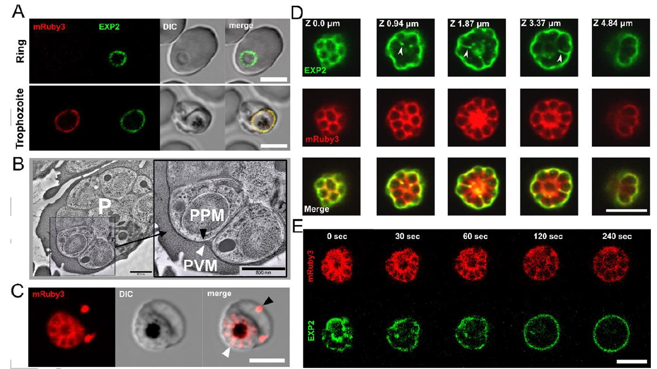
Visualization of parasitophorous vacuolar lumen and vacuolar membrane by light and electron microscopy. A. Visualization of the parasitophorous vacuole by detecting fluorescently tagged PVM-resident protein Exp2-mNeonGreen (green color) and soluble vacuolar fluorescent protein mRuby3 (red color) using live cell fluorescence microscopy. Note that the de novo produced mRuby3 peptide became visible starting from the trophozoite stage of parasite development, while Exp2 could be visualized started from the ring stage due to secretion from dense granules immediately following invasion. Both signals are co-localized in the trophozoite. B. EM images of schizont showing peripheral location of PVM during schizogony. PVM, (white arrowhead) and parasite plasma membrane (PPM, black arrowhead). P, parasite. Cryo-microscopy. Scale bar = 800 nm. C. The pattern of soluble PV-targeted mRuby3 protein distribution in the schizont stage: signal delineates de novo formed parasites arranged in a flower-like structure and marks both the PV compartment (white arrowhead) and the extra-parasitic membrane compartments in the erythrocyte cytoplasm (black arrowhead). D. A partial co-localization (white arrowhead) of vacuolar membrane and lumen fluorescent signals revealed in the optical sections of a schizont. Airy scan confocal microscopy. E. Photodamage and swelling of an infected erythrocyte leads to the redistribution of Exp2-mNeonGreen signal to the vacuolar periphery. Time 0: laser wounding of the erythrocyte using 800 nm two-photon illumination of the area outside the schizont location. All scale bars in the light-microscopy images are 5 mm.
Glushakova S, Beck JR, Garten M, Busse BL, Nasamu AS, Tenkova-Heuser T, Heuser J, Goldberg D, Zimmerberg J. Rounding precedes rupture and breakdown of vacuolar membranes minutes before malaria parasite egress from erythrocytes. Cell Microbiol. 2018 Jun