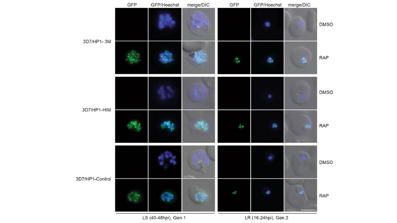
Sub-nuclear localization of PfHP1 phosphomutants. Representative live cell fluorescence images showing the localization of the GFP-tagged PfHP1–3M and PfHP1-HIM phosphomutants and the PfHP1-Control protein in late schizonts (LS, 40–48 hpi, generation 1; 40 hrs after rapamycin treatment) and after reinvasion in the progeny at late ring stage (LR, 16–24 hpi, generation 2; 64 hrs after rapamycin treatment). Nuclei were stained with Hoechst. DIC, differential interference contrast. Scale bar, 5 μm. Live cell fluorescence imaging in late schizonts at 40–48 hpi and in the late ring stage progeny at 16–24 hpi in generation 2 showed that the GFP-tagged PfHP1–3M, PfHP1-HIM and control PfHP1-GFP were not expressed in DMSO-treated parasites as expected. However, in the rapamycin-treated populations the GFP-tagged PfHP1–3M and PfHP1-HIM phosphomutants were expressed and showed a punctate pattern at the nuclear periphery indistinguishable from that observed for the PfHP1-GFP control protein. These results demonstrate that the phosphorylation of serine residues in the PfHP1 hinge domain is not required for the correct targeting and localization of PfHP1 to heterochromatin.
Bui HTN, Niederwieser I, Bird MJ, Dai W, Brancucci NMB, Moes S, Jenoe P, Lucet IS, Doerig C, Voss TS. Mapping and functional analysis of heterochromatin protein 1 phosphorylation in the malaria parasite Plasmodium falciparum. Sci Rep. 2019 9(1):16720. PMID: 31723180