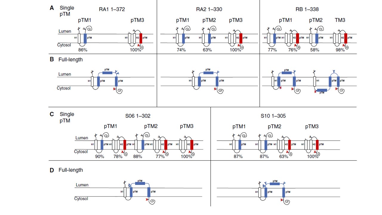
Schematic drawing representing the most common topologies of single putative transmembrane (pTM) domains or full-length RIFIN and STEVOR proteins using Lep model proteins. Topologies of RA1, RA2, and RB with single pTM domains (A) and full-length (B). Topologies of S06 and S10 single pTM domains (C) and full-length (D). In all cases, the model protein Lep was used; the fragment inserted in position LepH2 is blue and in LepH3 is red. GS is indicated with (Y) and unglycosylated sites indicated with a red (X) superimposed on the GS.
Andersson A, Kudva R, Magoulopoulou A, Lejarre Q, Lara P, Xu P, Goel S, Pissi J, Ru X, Hessa T, Wahlgren M, von Heijne G, Nilsson I, Tellgren-Roth Å. Membrane integration and topology of RIFIN and STEVOR proteins of the Plasmodium falciparum parasite. FEBS J. 2019 Dec 10.
Other associated proteins
| PFID | Formal Annotation |
|---|---|
| STEVOR | STEVOR |