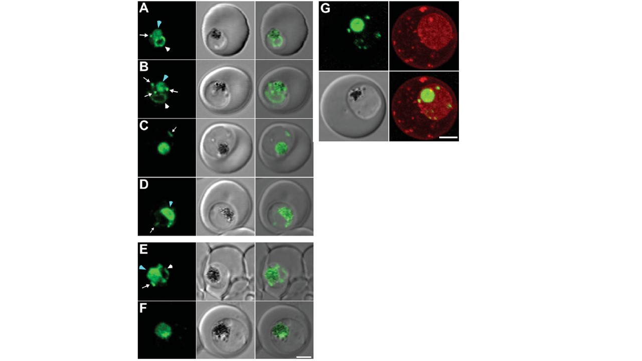
Expression of PM2–GFP at different stages of the intra-RBC cycle of P. falciparum transfectants (A–D) 3D7 and (E, F) Dd2 transfectants expressing PM2–GFP were imaged at the young to mid trophozoite (A, B, E) or mature trophozoite to schizont (C, D, F) stage. The images (left to right) represent a GFP fluorescence image, a DIC (differential interference contrast) image and an overlay of these images. DV-located GFP is indicated with a blue arrowhead. ER-located GFP is indicated with white arrowheads, while cytostomal vesicles are indicated with white arrows. (G) A 3D7-PM2 trophozoite was labelled with BODIPY® ceramide and the GFP (green fluorescence) and BODIPY® (red fluorescence) signals were imaged by confocal microscopy. The images shown represent an average projection obtained from a series of optical sections. Note: the intensities of the images were adjusted to optimize the fluorescence signal at each parasite stage. Scale bar, 2 μm.
Klonis N, Tan O, Jackson K, Goldberg D, Klemba M, Tilley L. Evaluation of pH during cytostomal endocytosis and vacuolar catabolism of haemoglobin in Plasmodium falciparum. Biochem J. 2007 407(3):343-54.