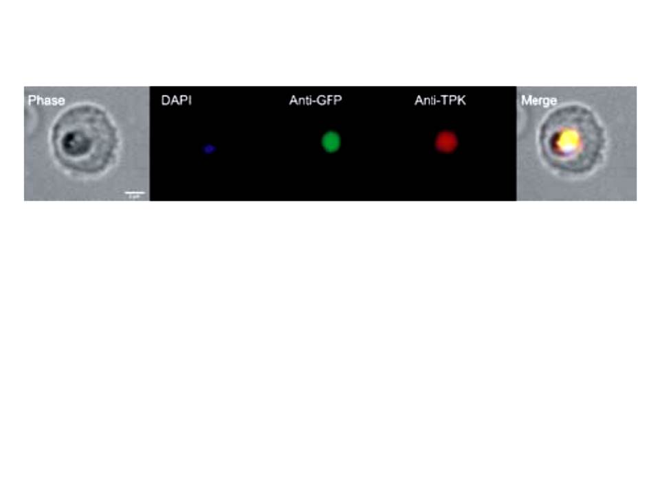
Immunofluorescence of plasmodial TPK. Immunofluorescence analyses were performed in 3D7 parasites expressing a cytosolic GFP reporter protein (Struck et al., 2005). The image shows single infection of an erythrocyte by P. falciparum (trophozoite stage), which was fixed onto a glass microscope slide and incubated with the DNA-specific dye DAPI (DAPI), a monoclonal anti-GFP antibody (Anti-GFP) and anti-PfTPK antibodies (Anti-PfTPK), respectively. The phase contrast image (Phase) was analysed in the merge image (Merge) revealing co-localisation of the GFP reporter and TPK protein – TPK is localized in the parasite’s cytoplasm.
Eschbach ML, Müller IB, Gilberger TW, Walter RD, Wrenger C. The human malaria parasite Plasmodium falciparum expresses an atypical N-terminally extended pyrophosphokinase with specificity for thiamine. Biol Chem. 2006 387:1583-91. Copyright Walter de Gruyter GmbH 2009.