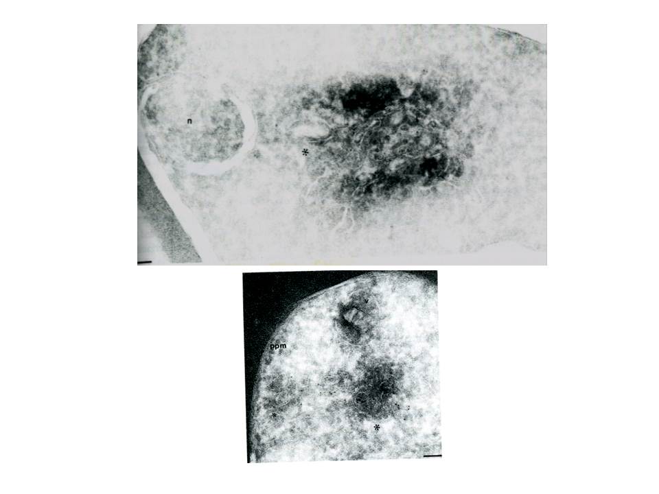
Cryosections of ring/trophozoite-infected Erythrocytes were probed with anti-Pf ERD2 (A) antibodies from non-immune sera (B) or anti-Pfrab6 (C) and goat anit-rabbit antibodies conjugated to 5 nm gold particles. In panel A, labeling of PfERD2 is detected in a perinuclear, tubulovesicular structure (*) with 1:100 anti PfERD2. The nucleus is marked by a n. Bar indicates 0.08 mm. In panel B, labeling of Pfrab6 (1:1OO) is seen in tubulovesicular clusters in peripheral regions of the cell. One cluster (large *) is proximal to the digestive food vacuole (marked with v). A second tubulovesicular cluster (small *) lies next to the parasite plasma membrane (marked by ppm). Bar indicates 0.1 mm.
Van Wye J, Ghori N, Webster P, Mitschler RR, Elmendorf HG, Haldar K. Identification and localization of rab6, separation of rab6 from ERD2 and implications for an 'unstacked' Golgi, in Plasmodium falciparum. Mol Biochem Parasitol. 1996 83:107-20. Copyright Elsevier 2010
Other associated proteins
| PFID | Formal Annotation |
|---|---|
| PF3D7_1144900 | ras-related protein Rab-6 |
