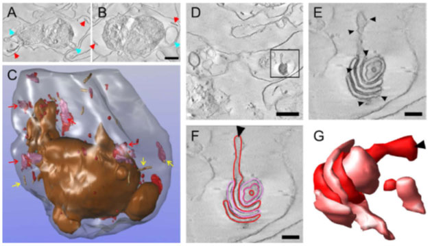
Three-dimensional (3-D) reconstruction of an entire Plasmodium falciparum strain D10-infected red blood cell (RBC) from electron tomograms of serial sections. Purified D10 parasite-infected RBCs were permeabilized with Equinatoxin II (Eqt II) and labelled with anti-membrane-associated histidine-rich protein-1 (MAHRP1), and immunogold protein A, fixed, stained, embedded in resin and sectioned (300 nm). Twenty-five serial sections were collected through a late trophozoite-infected RBC. (A and B) Selected virtual sections (12 nm) with Maurer’s clefts and tubulovesicular network (TVN) indicated with red and blue arrowheads. Scale bar, 1 lm. (C) Individual structures were segmented. The RBC membrane is rendered in translucent grey and the parasitophorous vacuole (PV) membrane and extensions from the PV membrane in bronze. Maurer’s cleft lamellae are depicted in red and mauve and are sometimes present as individual lamellae but often present as stacks. (Some examples are indicated with red arrows). Tubular structures are shown as beige cylinders (yellow arrows). (D) Virtual section (within the 15th physical section) with an individual Maurer’s cleft stack marked with a rectangle. Scale bar, 1 lm. (E) Virtual section of the same Maurer’s cleft stack from tomogram data collected at higher magnification. The presence of the gold particles (arrowheads) indicates labelling of the Maurer’s cleft resident, MAHRP1. This Maurer’s cleft stack spanned three sections. (F and G) Different lamellae of the Maurer’s clefts are indicated in red and pink in a virtual section and a rendered image. The arrowheads indicate the same extended region of the Maurer’s cleft in C and D. Scale bars, 200 nm.
Hanssen E, Carlton P, Deed S, Klonis N, Sedat J, Derisi J, Tilley L. Whole cell imaging reveals novel modular features of the exomembrane system of the malaria parasite, Plasmodium falciparum. Int J Parasitol. 2010 Jan;40(1):123-134. Copyright Elsevier 2010.