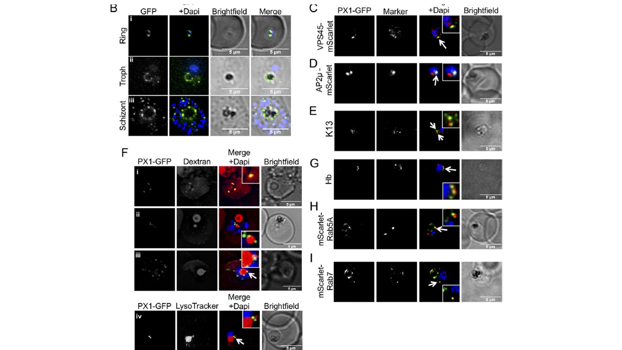
Subcellular localization of PfPX1-GFP. (A) Anti-GFP western blot of mixed stage PfPX1-GFP parasites showing the expression of the fusion protein. (B) Live cell imaging of the PfPX1-GFP parasite line at 3 different stages. Troph: trophozoite. (C) Colocalization of PfPX1-GFP with the endolysosomal marker PfVPS45-mScarlet. White arrow shows the overlapping foci. (D) Colocalization of PfPX1-GFP with PfAP2m-mScarlet. White arrow shows the overlapping foci. (E) Colocalization of PfPX1-GFP with PfKelch13 (K13) using an anti-PfK13 antibody. White arrow shows the overlapping foci. (F) PfPX1-GFP in red blood cells preloaded with dextran Alexa-594. (F, iv) Colocalization of PfPX1-GFP with LysoTracker. White arrow in iii and iv show the DV extension. Single optical slices shown. (G) Colocalization of PfPX1-GFP with hemoglobin-filled structures. White arrow shows the overlapping foci. (H) Colocalization of PfPX1-GFP with mScarlet-PfRab5a. White arrow shows the juxtaposed foci. (I) Colocalization of PfPX1-GFP with mScarlet-PfRab7. White arrow shows the juxtaposed foci. Dapi: parasite nuclei.
Mukherjee A, Crochetière MÈ, Sergerie A, Amiar S, Thompson LA, Ebrahimzadeh Z, Gagnon D, Lauruol F, Bourgeois A, Galaup T, Roucheray S, Hallée S, Padmanabhan PK, Stahelin RV, Dacks JB, Richard D. A Phosphoinositide-Binding Protein Acts in the Trafficking Pathway of Hemoglobin in the Malaria Parasite Plasmodium falciparum. mBio. 2022 Jan 18;13(1):e0323921. doi: 10.1128/mbio.03239-21. Epub ahead of print. PMID:
Other associated proteins
| PFID | Formal Annotation |
|---|---|
| PF3D7_0211200 | ras-related protein Rab-5A |
| PF3D7_0216400 | vacuolar protein sorting-associated protein 45, putative |
| PF3D7_0903200 | ras-related protein RAB7 |
| PF3D7_1218300 | AP-2 complex subunit mu |
| PF3D7_1343700 | kelch protein K13 |
