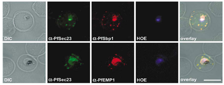
Double indirect immunofluorescence microscopy. Co-localization of PfSec23 with PfSbp1 PFE0065w (first row) and with PfEMP1 Ab to the cytoplasmic domain (second row). Infected erythrocytes were fixed in ice cold acetone/10% methanol for 5 min, blocked in PBS containing 5% skim milk powder and then incubated for 120 min at room temperature with the primary antibodies. Antibodies were diluted as follows: guinea pig antibodies against PfEMP1 (1:200), mouse antibodies against PfSec23 (1:20) and PfSbp1 (1:100). Secondary antibodies (Dianova, Germany) were either conjugated to Cy3 (anti-mouse antibodies) or to FITC (anti-guinea pig and anti-mouse antibodies) and diluted as recommended by the supplier. Double immunofluorescence of anti-PfSec23 and anti-PfSbp1 was done sequentially. The nuclei of parasites were visualized by staining the parasite DNA with Hoechst 333258 (HOE). Fluorescence was detected using an LSM 510 confocal laser scanning microscope (Zeiss). Overlay: overlays of the three fluorescence micrographs. Bar: 7 mm. PfSec23 is localized in the parasite cytoplasm, as one would expect for a COPII protein present on vesicles that mediate anterograde protein transport from the ER to the Golgi. Interestingly, PfSec23 is also located in the host erythrocyte cytosol, where it co-localizes with PfEMP1 and PfSbp1 in Maurer’s clefts.
Wickert H, Rohrbach P, Scherer SJ, Krohne G, Lanzer M. A putative Sec23 homologue of Plasmodium falciparum is located in Maurer's clefts. Mol Biochem Parasitol. 2003 129:209-13. Copyright Elsevier 2009.