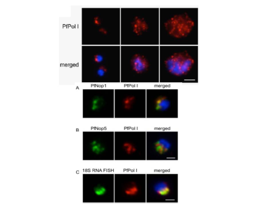
Upper 2 rows: Immunofluorescence analysis of PfPol I (red). Ring-stage parasites show a half-moon perinuclear nucleolus. In trophozoites, PfPol I shows an additional diffuse pattern in the nucleoplasm and cytoplasm. In schizont segmented forms, PfPol I appears to relocalize at the nuclear periphery. Parasite nuclei are shown by DAPI staining (blue). (Scale bars, 2 μm.)
Lower 3 rows: Double-labeling immunofluorescence assays using rabbit anti-PfPol I and rat anti-PfNop1 (A) or anti-PfNop5 (B) antibodies, showing localization of the nucleolus at periphery of nucleus, forming a “hat-like” structure. (C) RNA FISH (using a 18S universal probe that hybridizes to all rRNAs produced in the parasite, green) combined with immunofluorescence microscopy (using the anti-PfPolI antibody, red), demonstrating that the region detected by the antibodies corresponds to the nucleolus of the parasite. In all images, parasites are in the ring stage, and the nuclear DNA is stained with DAPI (blue). (Scale bars, 1 μm.)
Mancio-Silva L, Zhang Q, Scheidig-Benatar C, Scherf A. Clustering of dispersed ribosomal DNA and its role in gene regulation and chromosome-end associations in malaria parasites. Proc Natl Acad Sci U S A. 2010 107:15117-22.
Other associated proteins
| PFID | Formal Annotation |
|---|---|
| PF3D7_0509400 | RNA polymerase I |
| PF3D7_1407100 | rRNA 2'-O-methyltransferase fibrillarin, putative |
