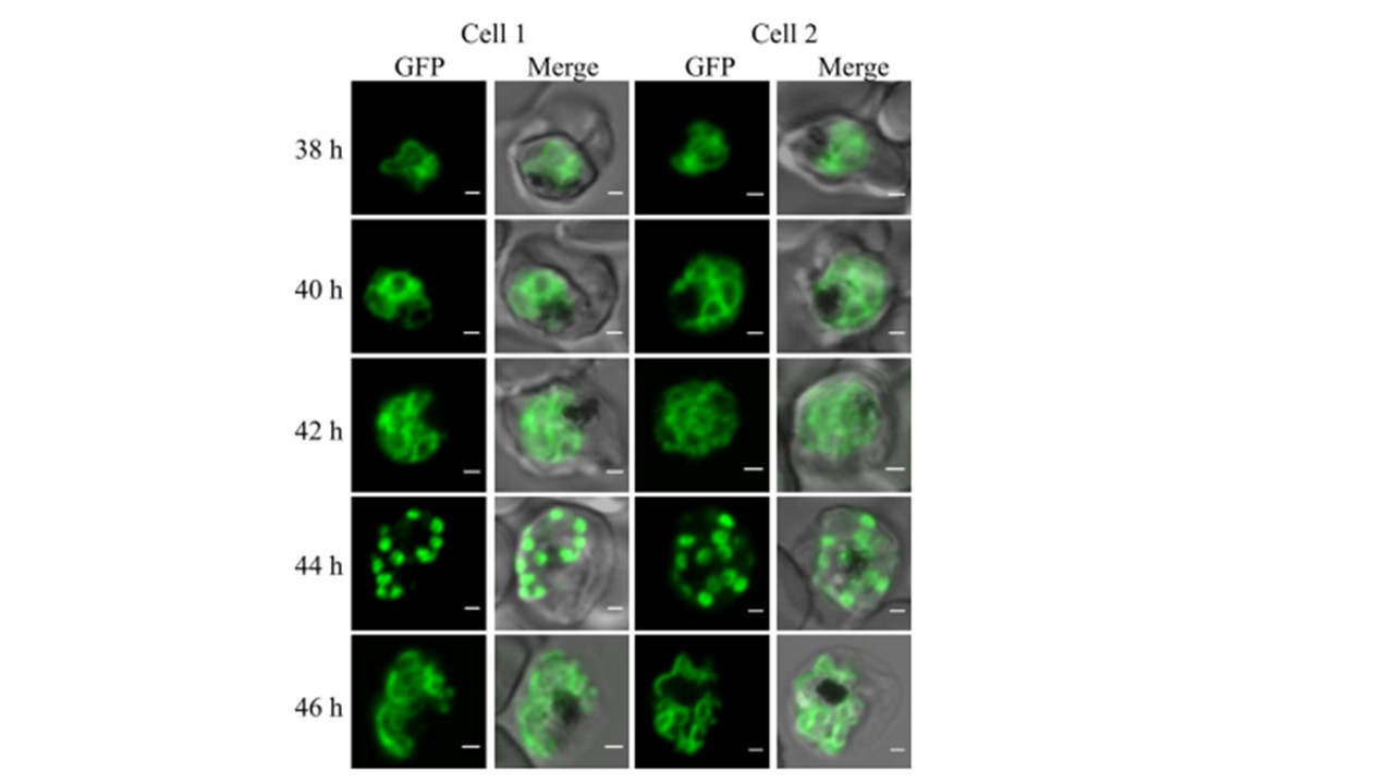
Dynamics of PfGAP50-GFP-labeled compartments in live transfectants. Shown is live confocal fluorescence microscopy of highly synchronized PfGAP50-GFP transfectants. Single section scans (collected at 25 s/pixel) from different cells at 2-h intervals in the schizont stage are shown (2 cells are represented per time point). At 40 to 42 h after invasion, reticular structures with looped extensions and focal concentrations of PfGAP50-GFP are apparent, coalescing into punctate structures by 44 h. The PfGAP50-GFP-containing structures then appear to expand around each of the daughter merozoites. Bars 1 mm.
PubMed Article: Tracking Glideosome-associated protein 50 reveals the development and organization of the inner membrane complex of Plasmodium falciparum
Other associated proteins
| PFID | Formal Annotation |
|---|---|
| PF3D7_0918000 | glideosome-associated protein 50 secreted acid phosphatase |
| PF3D7_1222700 | glideosome-associated protein 45 |