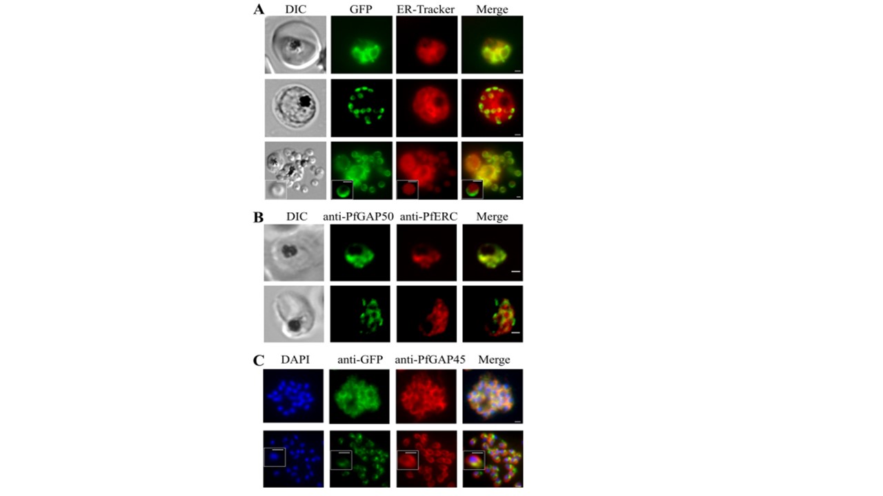
PfGAP50 is located in the ER prior to recruitment to the IMC. (A) Parasites expressing PfGAP50-GFP (green) were incubated with ER-Tracker (depicted in red). The GFP and ER-Tracker fluorescence signals are colocated in trophozoite stage parasites. Upon relocation of GFP fluorescence to the apical ends of nascent merozoites during the early schizont stage, the reticular ER-Tracker labeling persists. PfGAP50-GFP is concentrated at one pole of released merozoites, while ER-Tracker labels internal structures. (B) Fixed-cell immunofluorescence microscopy of the 3D7 parent strain labeled with antibodies raised against PfGAP50 (green) and PfERC (red). (C) Immunofluorescence microscopy of PfGAP50-GFP transfectants at the mature- and ruptured-schizont stages labeled with anti-GFP (green), anti-PfGAP45 (red), and DAPI (blue). Bars 1 mm.
Other associated proteins
| PFID | Formal Annotation |
|---|---|
| PF3D7_0918000 | glideosome-associated protein 50 secreted acid phosphatase |
| PF3D7_1222700 | glideosome-associated protein 45 |