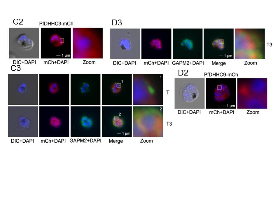
Over-expression and localization of PfDHHC3 and PFDHHC9 (PF11_0167) in late stage parasites. C. Expression of PfDHHC3-mCherry. C. Expression of PfDHHC3-mCherry. C2. Microscopic analysis located this GFP-fusion protein in the periphery of the nascent merozoites. C3. Co-localization with the IMC marker GAPM2 (anti-GAPM2, green) in fixed cells shows differential localization of PfDHHC2-mCherry with the IMC localization in early stages (T1) and colocalization in nascent merozoites (T3) consistent with plasma membrane association. Scale bar, 1 μm. D.
Expression of PfDHHC9-mCherry. D2. Microscopy localized this fusion protein mainly in apical structures in the parasite. D3. Co-localization with the IMC marker GAPM2 (anti-GAPM2, green) in fixed cells shows differential localization of PfDHHC9 with the IMC (T3). Scale bar, 1 μm.
Wetzel J, Herrmann S, Swapna LS, Prusty D, Peter AT, Kono M, Saini S, Nellimarla S, Wong TW, Wilcke L, Ramsay O, Cabrera A, Biller L, Heincke D, Mossman K, Spielmann T, Ungermann C, Parkinson J, Gilberger TW. The role of palmitoylation for protein recruitment to the inner membrane complex of the malaria parasite. J Biol Chem. 2014 Nov 25.
Other associated proteins
| PFID | Formal Annotation |
|---|---|
| PF3D7_0423500 | glideosome associated protein with multiple membrane spans 2 |
| PF3D7_1115900 | palmitoyltransferase DHHC9 |
