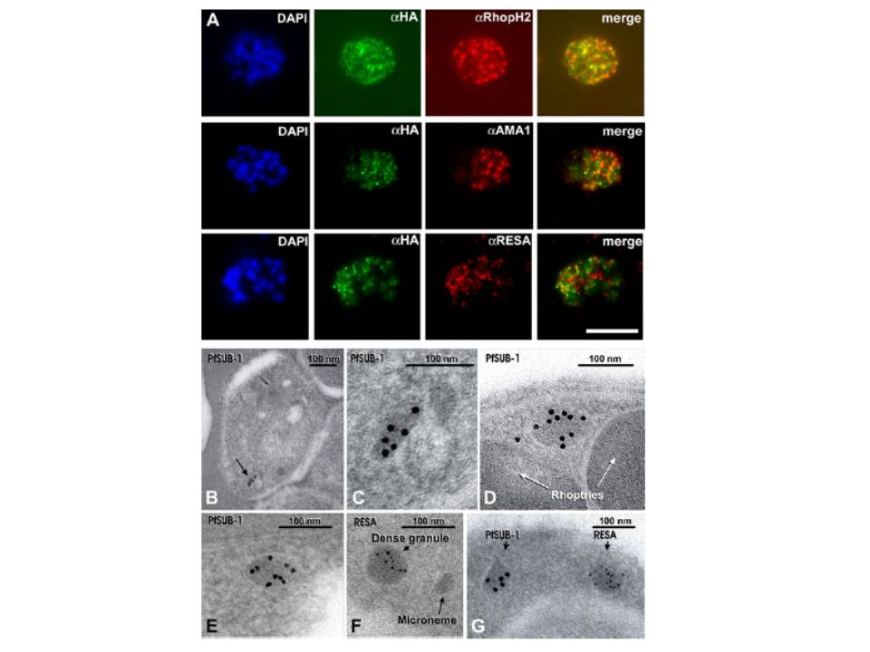
(A) Schizonts of 3D7SUB1HA3 clone C10 dual-labeled with the anti-HA mAb 3F10 (aHA; green) plus mAb 61.3 (aRhopH2), mAb 4G2 (aPfAMA1; micronemes, or mAb 28/2 (aRESA; dense granules. Nuclei were stained with DAPI (blue). Merged images (no DAPI) show that PfSUB1 localizes with none of the other markers. The scale bar represents 5 mm. Identical results were obtained with 3D7SUB1HA3 clone F7 or by using mAbs specific for RAP2 or EBA-175 as markers for rhoptries and micronemes respectively (data not shown). (B–G) Immunoelectron microscopic localization of PfSUB1 in P. falciparum schizonts. (B and C) Labeling of an elongated organelle, arrowed in (B) and at higher magnification in (C), by anti-PfSUB1 antibodies labeled with 10 nm immunogold. (D and E) Two more examples of PfSUB1 positive organelles are shown, also labeled with 10 nm immunogold. These are less elongate than in (B) and (C), reflecting some variation in organelle shape; however, they are typically ellipsoidal, in contrast with the rounded dense granules. (F and G) Staining for RESA with mAb 28/2 and 5 nm immunogold. In (F), an unlabeled microneme is also shown for size comparison. In (G) double staining for PfSUB1 and RESA with different sizes of immunogold shows that the two proteins are in different organelles.
Yeoh S, O'Donnell RA, Koussis K, Dluzewski AR, Ansell KH, Osborne SA, Hackett F, Withers-Martinez C, Mitchell GH, Bannister LH, Bryans JS, Kettleborough CA, Blackman MJ. Subcellular discharge of a serine protease mediates release of invasive malaria parasites from host erythrocytes. Cell. 2007 131:1072-83.
Other associated proteins
| PFID | Formal Annotation |
|---|---|
| PF3D7_0102200 | ring-infected erythrocyte surface antigen |
| PF3D7_0507500 | subtilisin-like protease 1 |
| PF3D7_0929400 | high molecular weight rhoptry protein 2 |
