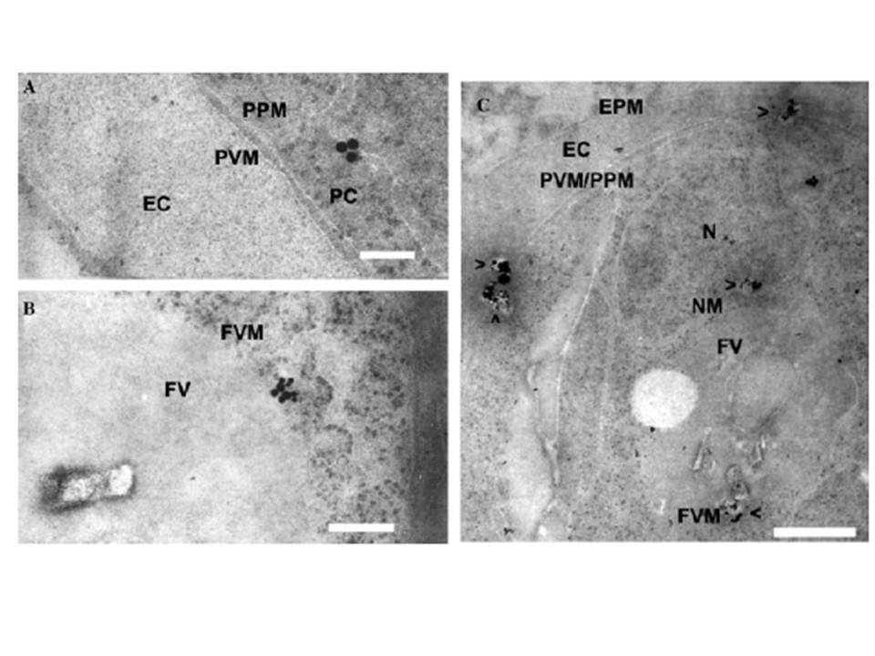
Location of Pfdyn1 in P. falciparum-infected erythrocytes with transmission electron. Percoll-synchonized late stage cells were fixed, and then incubated with purified anti-Pfdyn1-C antibody (1:100). Then incubated for in 15nm gold labeled goat anti-rabbit IgG diluted 1:20 in PBT and stained with uranyl acetate and lead citrate. FV, food vacuole; FVM, food vacuole membrane; EC, erythrocyte cytoplasm; EPM, erythrocyte plasma membrane; PC, parasite cytoplasm; PVM, parasitophorous vacuole membrane; PPM, parasite plasma membrane; N, nuclear; and NM, nuclear membrane. Bar, 100nm in (A), 200nm in (B), and 500nm in (C). Vesicles containing Pfdyn1-associated vesicles are indicated by arrowheads (>). The immunogold particles were observed in the cytoplasm of parasite and associated with membrane-like organelles such as food vacuoles, nucleus membrane, and some other unconfirmed membrane structures. Those particles were also seen in the cytoplasm of erythrocyte and on the parasite plasma membrane (PPM), parasitophorous vacuole membrane (PVM), and even erythrocyte plasma membrane (EPM).
Li H, Han Z, Lu Y, Lin Y, Zhang L, Wu Y, Wang H. Isolation and functional characterization of a dynamin-like gene from Plasmodium falciparum. Biochem Biophys Res Commun. 2004 320:664-71. Copyright Elsevier 2011
