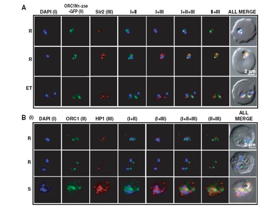
Co-localization of ORC1N1–238–GFP and Sir2 and immunolocalization of ORC1 and HP1 in 3D7. Glass slides containing the parasite smears from ORC1N1–238–GFP expressing parasites or 3D7 wild-type parasite lines were treated for immunolocalization using respective antibodies as indicated in the panels (A) and (B). (A) Co-localization of GFP and Sir2 in ORC1N1–238–GFP parasites during early stages of development as indicated on the left. ‘R’ indicates ring stage and ‘ET’ indicates early trophozoite stage parasites. DAPI shows the nuclei. Both ORC1 and Sir2 are localized to the nuclear periphery during the ring or early trophozoite stage. However, both proteins reorganize themselves at the onset of DNA replication and spread in the nucleus and cytoplasm. B(i): Both ORC1 and HP1 show nuclear punctate staining in 3D7 wild-type parasites during the early stages of development. ‘S’ indicates schizont stage parasites.
Deshmukh AS, Srivastava S, Herrmann S, Gupta A, Mitra P, Gilberger TW, Dhar SK. The role of N-terminus of Plasmodium falciparum ORC1 in telomeric localization and var gene silencing. Nucleic Acids Res. 2012 40(12):5313-31
Other associated proteins
| PFID | Formal Annotation |
|---|---|
| PF3D7_1203000 | origin recognition complex subunit 1 |
| PF3D7_1220900 | heterochromatin protein 1 |
