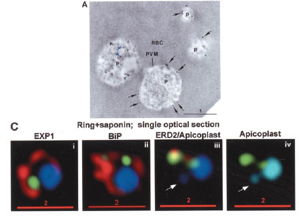
PTS-GFP is detected in the parasitophorous vacuole and in apposition with the Golgi in early rings. A, immunoelectron micrograph of rings showing localization of PTS (plastid-targeting-signal)-GFP in the parasitophorous vacuole and PVM (black arrows) as well as within the parasite (P; blue arrowheads). RBC, red blood cell. Scale bar, 1 mm.. C, i–iv: single optical sections showing green fluorescence in young rings permeabilized with 0.01% saponin relative to secretory markers PfEXP1, PfBiP, PfERD2 (shown in red in i–iii), and apicoplast DNA (marked with an arrow in iii and iv), as detected by indirect immunofluorescence and DeltaVision Microscopy. In C, iv, the Hoechst stain is pseudo-colored cyan to facilitate visualization of apicoplast DNA.
Cheresh P, Harrison T, Fujioka H, Haldar K. Targeting the malarial plastid via the parasitophorous vacuole. J Biol Chem. 2002 277:16265-77.
Other associated proteins
| PFID | Formal Annotation |
|---|---|
| PF3D7_0917900 | PfHsp70-2 |
| PF3D7_1121600 | parasitophorous vacuole membrane antigen QF 116 exported protein 1 circumsporozoite-related antigen |