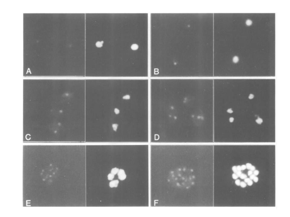
The identification of PfERD2 as a single discrete locus within the parasite. Thin blood smears were made from highly synchronized P.falciparum cultures, fixed and permeabilized, and incubated in affinity-purified PfERD2 antibody followed by incubation in FITC-conjugated goat anti-rabbit IgG (left panels). Nuclei were delineated by inclusion of 2 Ag/ml Hoechst in a final wash (right panels). Early ring stage infected erythrocytes, 10 h post-invasion (A); late ring stage infected erythrocytes, 22 h post-invasion (B); late trophozoite stage parasites, 34 h post-invasion (C and D); schizont stage parasite developing from four to eight nuclei (E); and late stage schizont, 16 nuclei (F). PfERD2 is tightly confined to a single focus of staining in the perinuclear region as seen by indirect immunofluorescence. This is redistributed by brefeldin A (BFA) to a diffuse pattern similar to that of parasite BiP, a marker for the ER; removal of the drug results in recovery of the single focus, consistent with the localization of PfERD2 to the parasite Golgi and its participation in a retrograde transport pathway to the ER
Elmendorf HG, Haldar K. Identification and localization of ERD2 in the malaria parasite Plasmodium falciparum: separation from sites of sphingomyelin synthesis and implications for organization of the Golgi. EMBO J. 1993 12(12):4763-73.