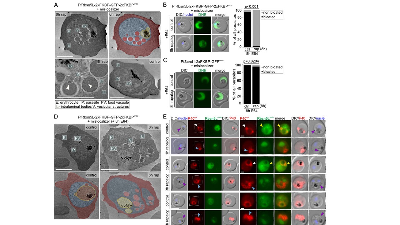
Vesicular structures after PfRbsn5L-inactivation are host cell cytosol uptake intermediates. (A) Electron microscopy images of PfRbsn5L knock-sideways (8 h rap) and control parasites. One representative image of n = 42 (rapalog) and 32 (control) cells. Top right shows the top left image with false coloring. E, erythrocyte (red); P, parasite (blue); FV; food vacuole (yellow); V; vesicular structure (red). White arrows indicate putative intraluminal bodies. Scale bars, 2 μm. (B, C) Live-cell images of PfRbsn5L (B) and PfSand1 (C) knock-sideways (rap+) and controls (ctrl) in parasites treated with E64 (E64+). Left: Live-cell images of representative DHE-stained parasites. Right: Quantification of number of cells with bloated FVs. Sabitzki R, Roßmann AL, Schmitt M, Flemming S, Guillén-Samander A, Behrens HM, Jonscher E, Höhn K, Fröhlke U, Spielmann T. Role of Rabenosyn-5 and Rab5b in host cell cytosol uptake reveals conservation of endosomal transport in malaria parasites. PLoS Biol. 2024 31;22(5):e3002639. PMID: 38820535
Other associated proteins
| PFID | Formal Annotation |
|---|---|
| PF3D7_1310300 | zinc finger protein, putative |