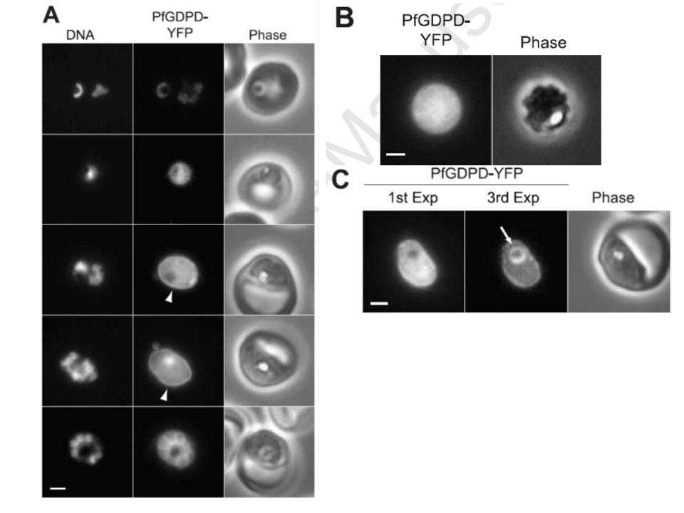
Localization of PfGDPD in asexual blood stage parasites. (A) Distribution of PfGDPD-YFP in live parasites at various stages of the asexual blood cycle. Parasites increase in maturity going from the top row (rings) to the bottom row (segmenting schizont). Rim fluorescence (PV) in mature trophozoites is indicated with an arrowhead. The exposure times and post acquisition processing parameters were identical for all YFP images to provide a sense of the relative expression levels of PfGDPD-YFP during the blood cycle. DNA was stained with Hoechst 33342. Bar, 2 mm. (B) Image of a parasite expressing PfGDPD-YFP that has been treated with 0.25 mg/mL saponin to permeabilize the erythrocyte and PV membranes. Bar, 2 mm. (C) The presence of YFP in the food vacuole is revealed after repeated exposures to excitation illumination. In the first one-second exposure (1st Exp), bright PV and cytosolic YFP fluorescence obscures the food vacuole fluorescence. Upon bleaching of PV and cytosolic YFP, the food vacuole fluorescence (arrow) becomes visible (3rd Exp). Bar, 2 mm.
Denloye T, Dalal S, Klemba M. Characterization of a glycerophosphodiesterase with an unusual tripartite distribution and an important role in the asexual blood stages of Plasmodium falciparum. Mol Biochem Parasitol. 2012 186(1):29-37