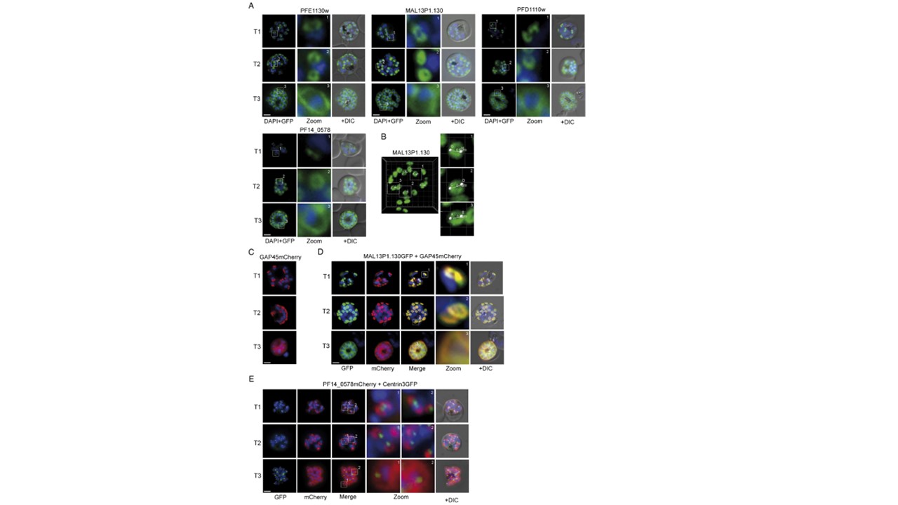
Group A–associated proteins colocalize with GAP45 during schizogony. (A) Using GFP as a fluorescence tag, the TMD proteins PFE1130w (PF3D7_0522600), MAl13P1.130 (PF3D7_1406800), PFD1110w (PF3D7_1323700), as well as PF14_0578 (PF3D7_1460600) reveal three distinct structures during ongoing schizogony. Commencing as cramplike structures (T1), they transform to small ring-shaped formations (T2) that toward the end of schizogony expand and are then equally distributed underneath the plasma membrane (T3). Enlargement of these distinct structures are marked (white square) and shown in second row (Zoom) for each cell line. (B) The ring structures highlighted by the TMD proteins and PF14_0578 have a maximum average diameter of
Kono M, Herrmann S, Loughran NB, Cabrera A, Engelberg K, Lehmann C, Sinha D, Prinz B, Ruch U, Heussler V, Spielmann T, Parkinson J, Gilberger TW. Evolution and architecture of the inner membrane complex in asexual and sexual stages of the malaria parasite. Mol Biol Evol. 2012 29(9):2113-32.
Other associated proteins
| PFID | Formal Annotation |
|---|---|
| PF3D7_0522600 | magnesium transporter NIPA, putative |
| PF3D7_1323700 | glideosome associated protein with multiple membrane spans 1 |
| PF3D7_1460600 | inner membrane complex sub-compartment protein 3 |
