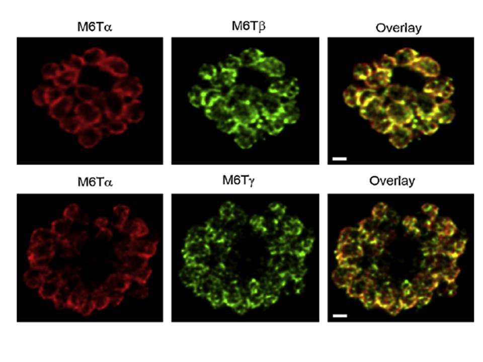
PfM6T proteins localize to the merozoite IMC. Confocal immunofluorescence images demonstrating location of each paralog, as indicated above each image. Images show a separate schizont-infected erythrocyte containing many merozoites; overlay of the red and green channels (rightmost column) shows colocalization of PfM6Ta with each other paralog. Red and green images reflect detection using specific antibodies raised in rabbits and mice, respectively. White scale bars represent 1mm.
Rayavara K, Rajapandi T, Wollenberg K, Kabat J, Fischer ER, Desai SA. A complex of three related membrane proteins is conserved on malarial merozoites. Mol Biochem Parasitol. 2009 167:135-43. Copyright Elsevier 2010
Other associated proteins
| PFID | Formal Annotation |
|---|---|
| PF3D7_0423500 | glideosome associated protein with multiple membrane spans 2 |
| PF3D7_1323700 | glideosome associated protein with multiple membrane spans 1 |
