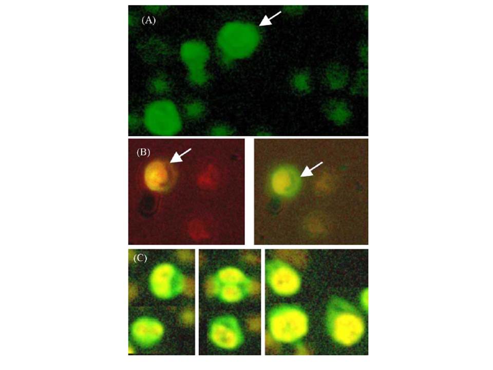
Confocal microscopy of parasitized erythrocytes detected using primary antibody generated with the recombinant pf CTP synthetase, and visualised with a secondary antibody labeled with FITC. (A) Parasite infected erythrocytes (indicated by an arrow) have a much brighter fluorescence compared to non-infected erythrocyte cells, which appear with a low level green auto-fluorescence. (B) Overlaid images from red and green wavelength of parasitized and non-parasitized erythrocytes. The arrows indicate the locations of pfCTP synthetase. (C) Parasitized erythrocytes at different stages of the parasite life cycle: ring stage, trophozoite stage and schizont stage (left to right).
Yuan P, Hendriks EF, Fernandez HR, O'Sullivan WJ, Stewart TS. Functional expression of the gene encoding cytidine triphosphate synthetase from Plasmodium falciparum which contains two novel sequences that are potential antimalarial targets. Mol Biochem Parasitol. 2005 143(2):200-8. PMID:
