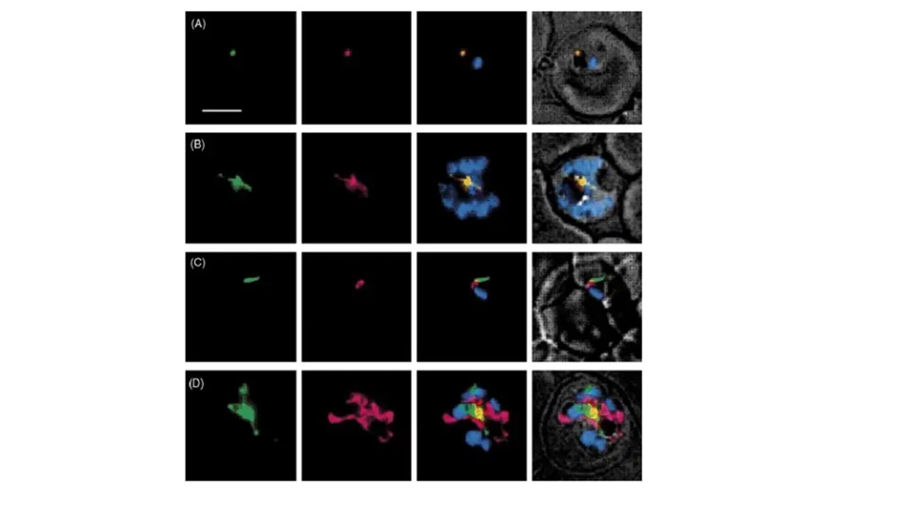
Subcellular localization of PfPREX. Malaria parasites were doubly transfected with pSSPF2/PfPREX-GFP and pSSPF2/Pf ACP-DsRED (A and B),or with pSSPF2/PfPREX-GFP alone (C and D). The mitochondrion of the pSSPF2/PfPREX-GFP single transfectant parasite (C and D) was stained with MitoTracker Red CM-H 2 XRos (MTR). Panels in each row represent: green signal of GFP (far left); red signal of DsRED specifically localizing to the apicoplast (A and B) or MTR to the mitochondrion (C and D) (second from the left); merge of the green and red signals as well as the blue signal of Hoechst33342 indicating the position of the parasite nucleus (second from the right); overlay of the merged image with the phase contrast image (far right). (A) Early trophozoite; (B) Schizont; (C) Early trophozoite; (D) Schizont. Scale bar: 5 mm.
Seow F, Sato S, Janssen CS, Riehle MO, Mukhopadhyay A, Phillips RS, Wilson RJ, Barrett MP. The plastidic DNA replication enzyme complex of Plasmodium falciparum. Mol Biochem Parasitol. 2005 141(2):145-153. PMID: 15926203.
