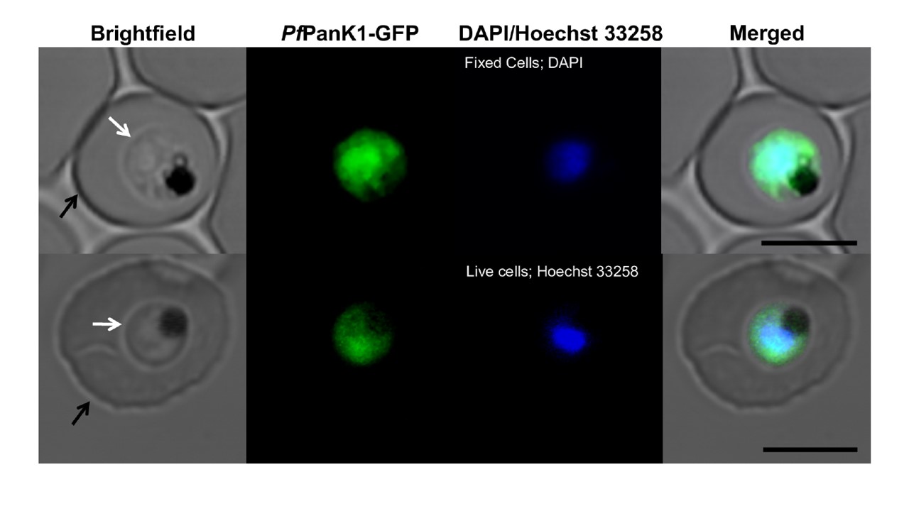
Confocal micrographs showing the subcellular location of GFP-tagged PfPanK1 in 3D7 strain parasites harbouring the Pfpank1- pGlux-1 episomal plasmid. From left to right: Brightfield, GFP-fluorescence, DAPI- (fixed cells; top) or Hoechst 33258- (live cells; bottom) fluorescence, and merged images of erythrocytes infected with trophozoite-stage P. falciparum parasites expressing PfPanK1-GFP. Arrows indicate the plasma membranes of either the erythrocytes (black) or the parasites (white). Scale bar represents 5 μm.
Tjhin ET, Spry C, Sewell AL, Hoegl A, Barnard L, Sexton AE, Siddiqui G, Howieson VM, Maier AG, Creek DJ, Strauss E, Marquez R, Auclair K, Saliba KJ. Mutations in the pantothenate kinase of Plasmodium falciparum confer diverse sensitivity profiles to antiplasmodial pantothenate analogues. PLoS Pathog. 2018 Apr 3;14(4):e1006918.
