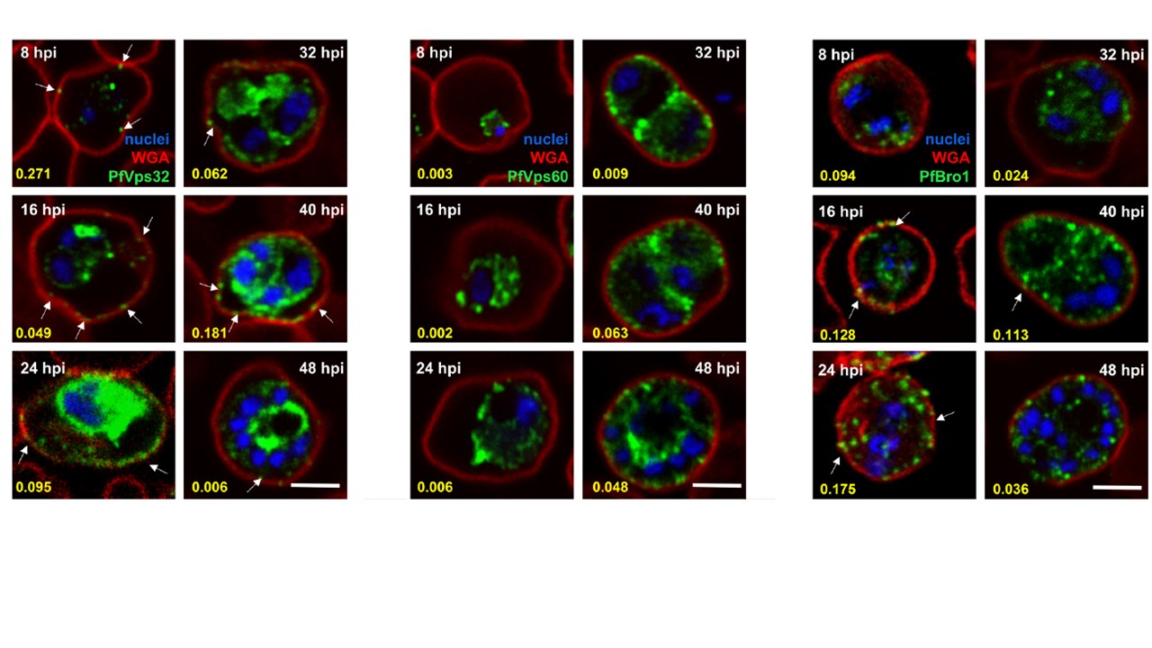
Expression and localization of PfVps32, PfVps60 and PfBro1 during the P. falciparum intraerythrocytic cycle. Human erythrocytes were infected with P. falciparum and fixed at different hpi. PfVps32, PfVps60 or PfBro1 (green) were detected by indirect confocal immunofluorescence microscopy and WGA (red) was used to label the RBC plasma membrane. Cell nuclei were visualized with Hoechst 33342 (blue). Arrows show protein-labeled puncta adjacent to the membrane of the pRBCs. Numbers in yellow indicate Manders’ overlap coefficients used to evaluate co-localization between WGA and each protein tested. Scale bar: 5 μm. Immunofluorescence assays showed that PfVps32, PfVps60 and PfBro1 were localized in the cytoplasm of the parasite, inside the parasitophorous vacuole (PV). In the case of PfVps32 and PfBro1, puncta stained with their respective antibodies were observed in the cytoplasm of parasitized erythrocytes outside the PV, some of them close to the RBC membrane. To examine whether these structures are exported to the parasitized RBC.
Avalos-Padilla Y, Georgiev VN, Lantero E, Pujals S, Verhoef R, N Borgheti-Cardoso L, Albertazzi L, Dimova R, Fernàndez-Busquets X. The ESCRT-III machinery participates in the production of extracellular vesicles and protein export during Plasmodium falciparum infection. PLoS Pathog. 2021 17(4):e1009455.
Other associated proteins
| PFID | Formal Annotation |
|---|---|
| PF3D7_1224200 | BRO1 domain-containing protein, putative |
| PF3D7_1243500 | vacuolar protein-sorting-associated protein 32, putative |