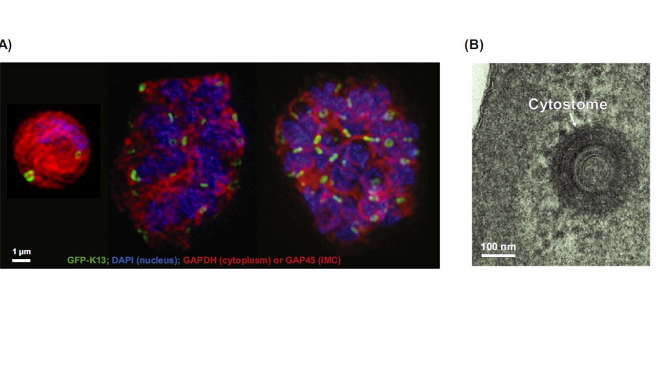
Kelch-13 (K13) Is Associated with Parasite Cytostomes. (A) Transfectants expressing GFP-tagged K13 at early trophozoite, early schizont, and segmented schizont stages, imaged using super-resolution light microscopy. K13, green; glyceraldehyde-3-phosphate dehydrogenase (PfGAPDH; cytoplasm) or glideosome-associated protein-45 (PfGAP45; inner membrane complex, IMC), red; 4′,6-diamidino-2-phenylindole (DAPI; nucleus), blue. Scale bar: 1 μm. The size, morphology, and cell location of the K13-labelled structure is consistent with a cytostome. (B) Electron micrograph showing a transverse section through a cytostomal ring with an external diameter of ~190 nm. Scale bar: 100 nm.
Yang T, Yeoh LM, Tutor MV, et al. Decreased K13 Abundance Reduces Hemoglobin Catabolism and Proteotoxic Stress, Underpinning Artemisinin Resistance. Cell Rep. 2019;29(9):2917–2928.e5.
r
Other associated proteins
| PFID | Formal Annotation |
|---|---|
| PF3D7_1222700 | glideosome-associated protein 45 |
| PF3D7_1343700 | kelch protein K13 |
