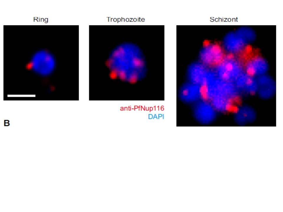
Nuclear pore redistribution throughout the intraerythrocytic stages. Immuno-fluorescence staining of nuclear pores using anti-PfNup116 at different intra-erythrocytic stages. Schizont stage image is a maximum ntensity projection. Nucleus stained by DAPI. Bar, 1 μm.
Guizetti J, Martins RM, Guadagnini S, Claes A, Scherf A. Nuclear pores and perinuclear expression sites of var and rDNA genes correspond to physically distinct regions in Plasmodium falciparum. Eukaryot Cell. 2013 12(5):697-702.
