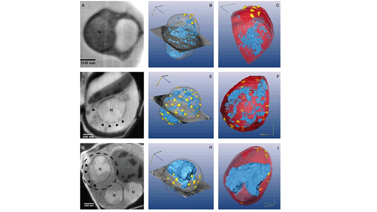
3D analysis of P. falciparum nuclear dynamics obtained by serial surface view imaging (FIB ‘slice and view’). Contrast is inverted with respect to TEM, so that the dense genetic matter is brighter and the loose genetic matter is relatively dark. A. A slice through ring stage nucleus. B. 3D model of the entire nucleus of a ring stage parasite derived from 134 slices at 10 nm resolution; grey – nuclear membrane; yellow – NPC; blue – heterochromatin. C. A cross-section through two plains of a model of the ring stage nucleus showing an inner view of the nucleus. Red – nuclear membrane; yellow – NPC; blue – heterochromatin. D. A slice through trophozoite nucleus. The nuclear pores are marked by arrowheads. E. 3D model of the entire trophozoite nucleus derived from 170 slices at 10 nm resolution; grey – nuclear membrane; yellow – NPC; blue – heterochromatin. F. A cross-section through two plains of a model of the trophozoite nucleus showing an inner view of the nucleus. Red – nuclear membrane; yellow – NPC; blue – heterochromatin. G. A slice through mid schizont nucleus. The nuclear pores are marked by arrowheads H. 3D model of the entire mid schizont nucleus derived from 136 slices; grey – nuclear membrane; yellow – NPC; blue – heterochromatin. I. A cross-section through two of the plains of a model of the mid schizont nucleus showing an inner view of the nucleus. Red – nuclear membrane; yellow – NPC; blue – heterochromatin.
Weiner A, Dahan-Pasternak N, Shimoni E, Shinder V, von Huth P, Elbaum M, Dzikowski R. 3D nuclear architecture reveals coupled cell cycle dynamics of chromatin and nuclear pores in the malaria parasite Plasmodium falciparum. Cell Microbiol. 2011 13(7):967-77. PMID: 21501361