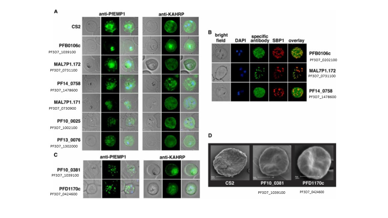
Microscopic Analysis of Mutants with Export Defects (A) Localization of PfEMP1 and KAHRP in mutant P. falciparum-infected erythrocytes. The parasite lines shown are those with either no PfEMP1 or reduced levels on the surface of infected erythrocytes determined by FACS and trypsin analysis. The first panel depicts localization of PfEMP1 and the second panel shows localization of KAHRP. The first column of each panel shows a bright-field image, the second panel specific antibody (either anti-PfEMP1 or anti-KAHRP) and the third panel overlay of the previous two and a nuclear stain (DAPI). (B) Localization pattern of three proteins which when deleted ablate surface exposure of PfEMP1. These proteins were detected with specific antibodies raised against the gene products of PFB0106c, MAL7P1.172 and PF14_0758 (see Figure C). The first panel shows a brightfield image, followed by a DAPI image, then the specific antibody (green), then antibodies against the Maurer's cleft resident protein SBP1 (red) and an overlay of the specific antibody with SBP1 localization. (C) Localization of PfEMP1 (first panel) and KAHRP (second panel) for parasite lines CS2ΔPF10_0381 and CS2ΔPFD1170c. Shown are a brightfield image, specific antibody (either anti-PfEMP1 or anti-KAHRP) and an overlay of the two with a nuclear stain (DAPI). Maier AG, Rug M, O'Neill MT, Brown M, Chakravorty S, Szestak T, Chesson J, Wu Y, Hughes K, Coppel RL, Newbold C, Beeson JG, Craig A, Crabb BS, Cowman AF. Exported proteins required for virulence and rigidity of Plasmodium falciparum-infected human erythrocytes. Cell. 2008.