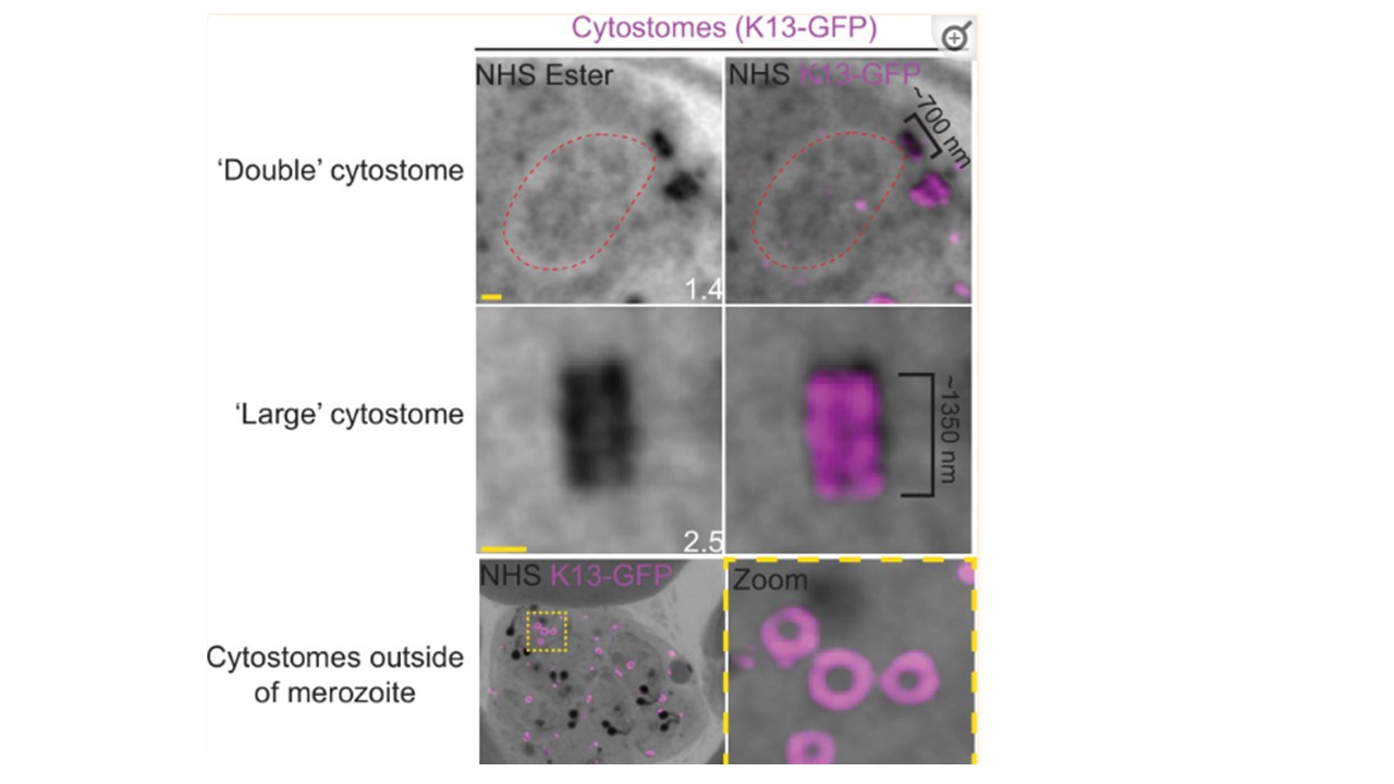
Observed cytostome morphologies.
Parasites expressing a GFP-tagged copy of the cytostome marker Kelch13 (K13-GFP) were prepared for ultrastructural expansion microscopy (U-ExM), stained with N-hydroxysuccinimide (NHS) ester (grayscale) and anti-GFP (cytostome; magenta) antibodies and imaged using Airyscan microscopy. ‘Double’ cytostomes where two collars appeared to be stacked on top of each other, and cytostomes approximately twice the diameter of other cytostomes were occasionally observed. Additionally, in almost all segmenting schizonts, multiple cytostomes were observed that were not incorporated into the forming merozoites. Images are maximum-intensity projections, number on image = Z-depth in µm of projection. Black scale bar = 2 µm, yellow scale bars = 500 nm. Liffner B, Cepeda Diaz AK, Blauwkamp J, Anaguano D, Frölich S, Muralidharan V, Wilson DW, Dvorin J, Absalon S. Atlas of Plasmodium falciparum intraerythrocytic development using expansion microscopy. 2023 9:2023.03.22.533773. PMID: 36993606