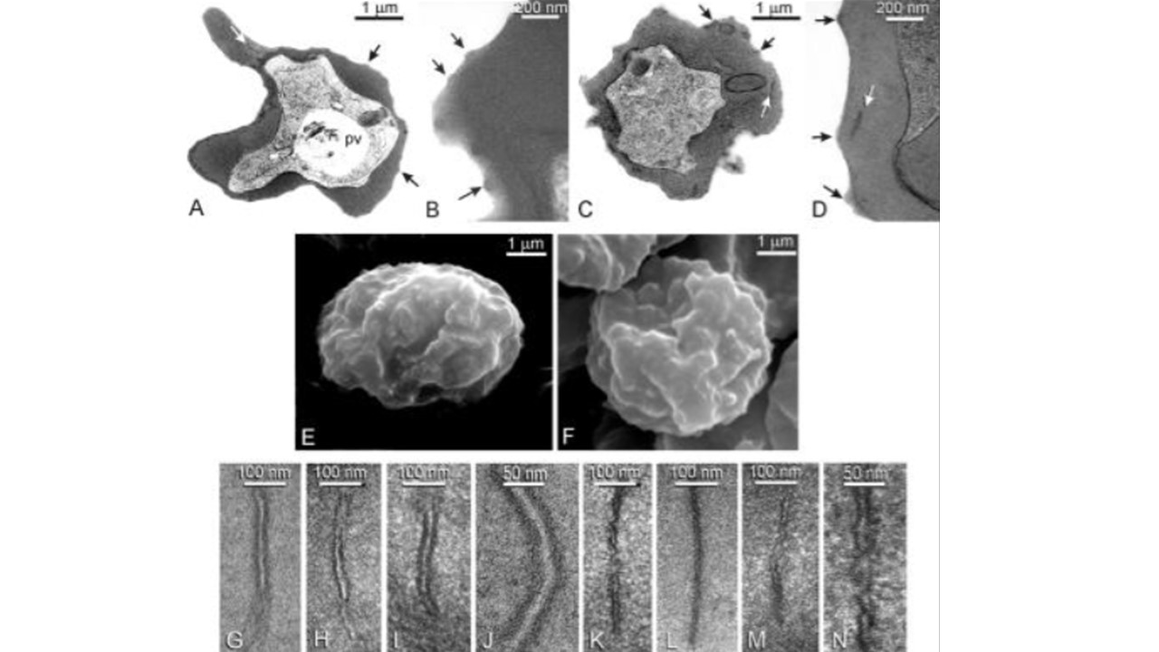
Ultrastructural analysis of IRBCs. Comparisons of Maurer's clefts and surface knob appearances in RBCs infected with wild-type 3D7 (A, B, E, and G–J) or sbp1-deleted 1G8 parasites (C, D, F, and K–N). Survey transmission electron micrographs of 3D7 (A and B) and 1G8 (C and D) show that both express Maurer's clefts (white arrows) and knobs (black arrows), and scanning electron microscopy of 3D7 (E) and 1G8 (F) also shows a similar expression of knobs. However, transmission electron microscopy at higher magnifications shows differences between 3D7 clefts (G–J) and those of 1G8 (K–N), in which clefts are typically narrower with a reduced intermembrane space; shown in higher magnification in J (3D7) and N (1G8). pv, pigment vacuole. Cooke BM, Buckingham DW, Glenister FK, Fernandez KM, Bannister LH, Marti M, Mohandas N, Coppel RL. A Maurer's cleft-associated protein is essential for expression of the major malaria virulence antigen on the surface of infected red blood cells. J Cell Biol. 2006. PMID: 16520384
=•Òµ