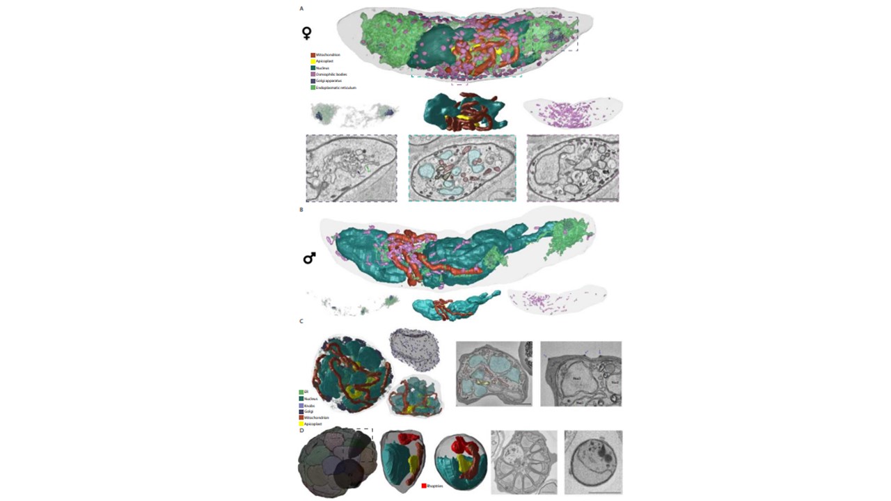
Ultrastructural features and renderings of mature gametocytes, schizont and segmented schizont. A Rendering of ultrastructural features of a mature
female gametocyte and specific renderings of Golgi and ER distribution (left panel), relation of nucleus/apicoplast/mitochondrion (middle panel) as well as distribution and appearance of osmiophilic bodies. Appearance of rendered structures in exemplar micrographs is matched to rendered features through color-coded dashed lines. Scale bars = 1 μm. B Rendering of ultrastructural features of a mature male gametocyte and specific renderings as in (A). Note the relative paucity of ER, Golgi, and osmiophilic bodies relative to the female gametocyte. C Rendering of ultrastructural features of an early schizont and specific rendering of relation of nucleus/apicoplast/mitochondrion as well as individual knob distribution. Appearance of ultrastructural features is shown in two exemplar micrographs. Scale bars = 1 μm. M Mitochondrion, ER Endoplasmic reticulum, Nucl Nucleus, FV Food vacuole. D Rendering of daughter merozoites within a segmented schizont and rendering of one intracellular and one extracellular merozoite with ultrastructural features. Overview on appearance of the two merozoite shapes is shown in two exemplar micrographs. Scale bars = 1 μm. For gametocytes all observations are based on and consistent throughout four biological replicates. The asexual blood stage observations are based on two biological replicates. Evers F, Roverts R, Boshoven C, Kea-Te Lindert M, Verhoef JMJ, Sommerdijk N, Sinden RE, Akiva A, Kooij TWA. Comparative 3D ultrastructure of Plasmodium falciparum gametocytes. Nat Commun. 2025