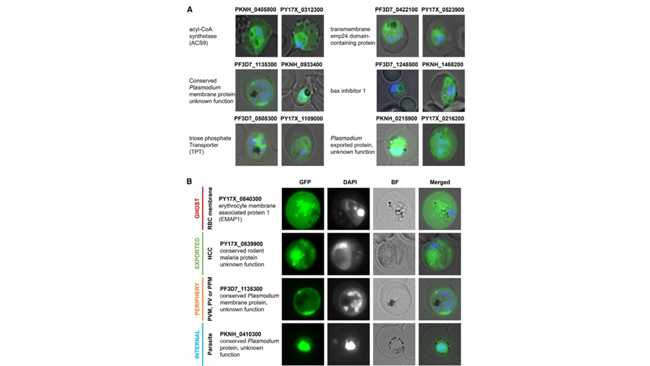
Live-cell fluorescence microscopy of GFP-tagged proteins. Representative images of P. falciparum-, P. knowlesi-, and P. yoelii-infected RBCs depicting (A) proteins from six orthology groups having localization data obtained from two parasite species and (B) the four localization patterns (ghost, exported, periphery, or internal) of the GFP-tagged protein (green), distinguished by microscopy imaging. The parasite nucleus was stained with DAPI (blue). Siau A, Ang JW, Sheriff O, Hoo R, Loh HP, Tay D, Huang X, Yam XY, Lai SK, Meng W, Julca I, Kwan SS, Mutwil M, Preiser PR. Comparative spatial proteomics of Plasmodium-infected erythrocytes. Cell Rep. 2023 42(11):113419. PMID: 37952150.