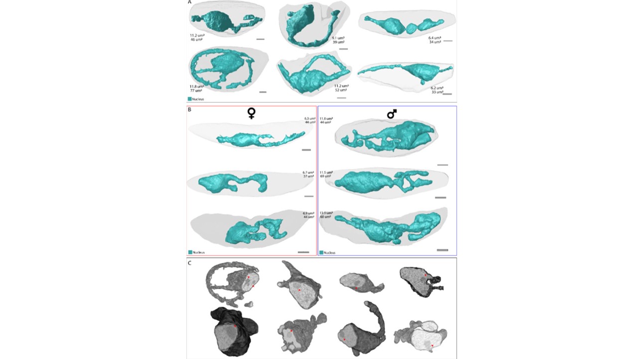
Distinct nuclear morphology in gametocytes. Renderings of nuclei (teal) in (A) stage II-III and (B) stage IV-V gametocytes. Stage IV-V gametocytes are divided in female (red outline) and male (blue outline) gametocytes based on the number and appearance of osmiophilic bodies, haemozoin distribution, and ER prevalence. For all nuclei, the respective volumes and surface areas are indicated. C Rendering of cross section of nuclei with grey values from EM data overlaid. Red arrowheads point at the position of the putative nucleolus. Evers F, Roverts R, Boshoven C, Kea-Te Lindert M, Verhoef JMJ, Sommerdijk N, Sinden RE, Akiva A, Kooij TWA. Comparative 3D ultrastructure of Plasmodium falciparum gametocytes. Nat Commun. 2025