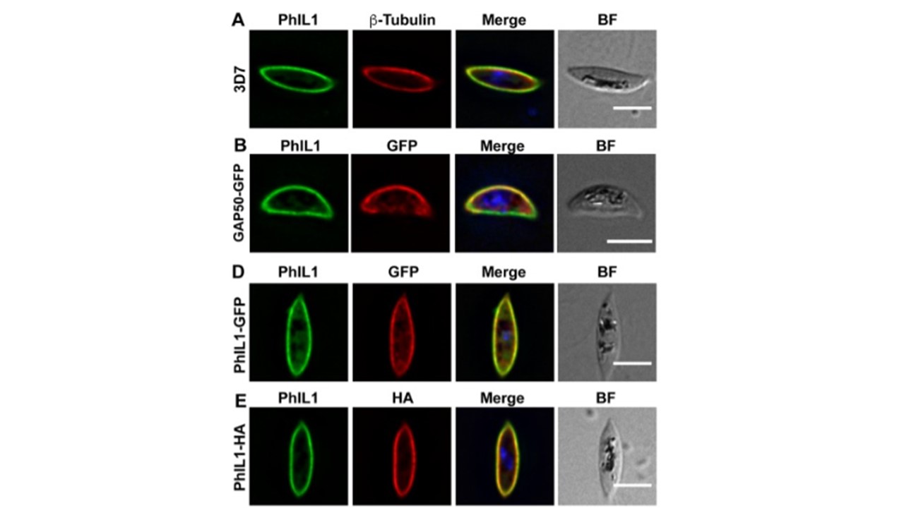
PhIL1 is located at the IMC in gametocytes. (A) Immunofluorescence microscopy showing anti-PhIL1 (green) at the periphery of a 3D7 stage IV gametocyte, showing fluorescence close to the anti-β-tubulin (red) labeling. (B) Immunofluorescence microscopy of a stage IV gametocyte showing overlap of GAP50-GFP (red) and PhIL1 (green) at the periphery of the cell. (D, E) Immunofluorescence microscopy of stage IV PhIL1-GFP (D) and PhIL1-HA (E) gametocyte transfectants. Parasites were labeled with anti-PhIL1 (green) and anti-GFP or anti-HA (red). Nuclei were labeled with DAPI. Scale bars: 5 μm. Parkyn Schneider M, Liu B, Glock P, Suttie A, McHugh E, Andrew D, Batinovic S, Williamson N, Hanssen E, McMillan P, Hliscs M, Tilley L, Dixon MWA. Disrupting assembly of the inner membrane complex blocks Plasmodium falciparum sexual stage development. PLoS Pathog. 2017 13(10):e1006659. PMID: 28985225
Other associated proteins
| PFID | Formal Annotation |
|---|---|
| PF3D7_1008700 | tubulin beta chain |