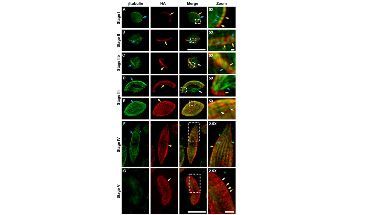
Genesis and development of the gametocyte IMC. (A-G)3D-SIM immunofluorescence microscopy reveals the location of PhIL1-HA (red) relative to β-tubulin (green) for PhIL1-HA gametocytes from stage I-V of development. (A) A row ofPhIL1-HAlabeled puncta(yellow arrow)is observed along one edge of the parasite, interleaved with β-tubulin staining (blue arrow). (B) In stage II gametocytes, PhIL1-HA is observed as 13ring-like structures (yellow arrows) aligned at the periphery of the parasites. Accumulations of β-tubulin staining can be seen in the centers of these disks (blue arrow). (C) The nascent IMC plates are more homogenously labeled with PhIL1-HA(yellow arrow), forming a ribbon-like structure at the periphery of the cell. Microtubules align with the IMC plates (blue arrow) or cross the parasite cytoplasm (white arrow). (D-E) In stage III, the IMC plates are clearly defined with homogenous PhIL1-HA labeling (yellow arrow). Bundles of microtubules underlie the IMC (blue arrow) or cross the parasite cytoplasm (white arrow). (F) In stage IV gametocytes, the IMC largely encases the parasite. The PhIL1-HAplates (yellow arrow) are still clearly visible and the microtubule network is tightly associated with the IMC (blue arrow). (G) By stage V, the microtubule network is disassembled but the IMC (labeled with PhIL1-HA) remains at the parasite periphery (yellow arrows; sutures are indicated with white arrows). 5x and 2.5X zoom images are shown on the right hand side. Scale bars: 5 μm. Scale bars: zoom images: 1μm.The represented area is marked on the images with a white box. He L, Qiu Y, Pang G, Li S, Wang J, Feng Y, Chen L, Zhu L, Liu Y, Cui L, Cao Y, Zhu X. Plasmodium falciparum GAP40 Plays an Essential Role in Merozoite Invasion and Gametocytogenesis. Microbiol Spectr. 2023 11(3):e0143423. PMID: 37249423
ÿ