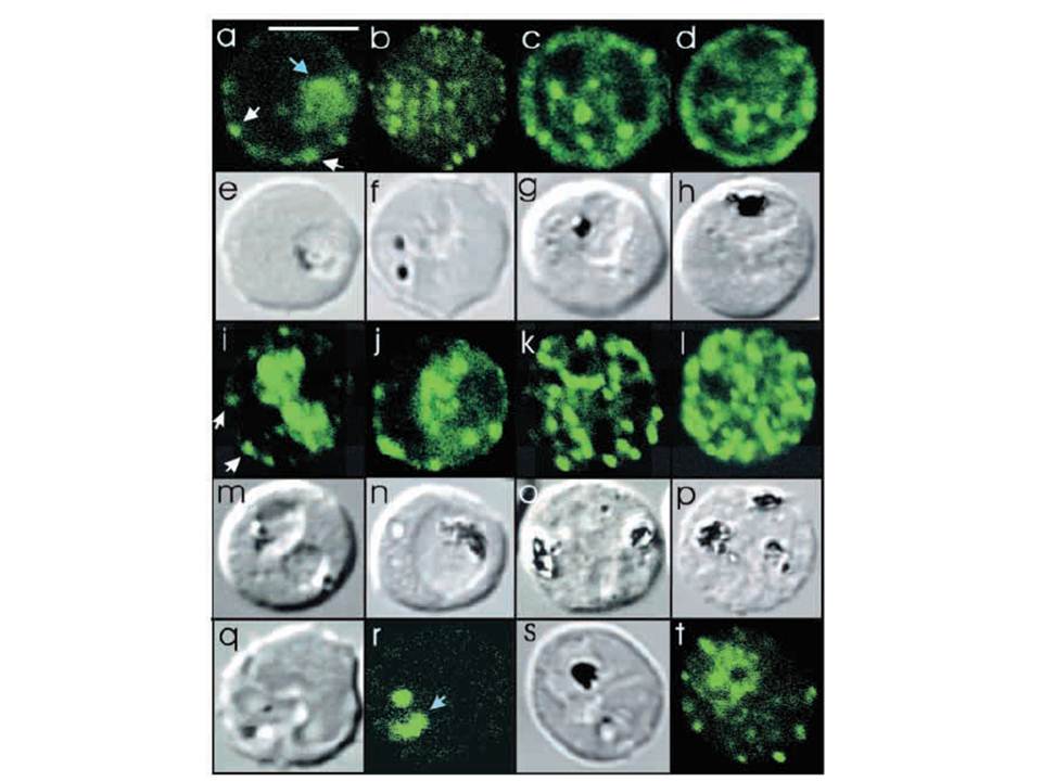
Intracellular location of PfSec31p and effect of BFA treatment on export of PfSec31p. Erythrocytes infected with ring stage (a,b,i) or mature (c,d,j-l) malaria parasites (K1 strain) were labelled with affinity purified rabbit anti-PfSec31(WD) antiserum (a-d) or affinity purified rabbit anti-PfSec31(int) antiserum (i-l) followed by a fluorescein-conjugated anti-rabbit IgG. Transmission images of a-d are shown in e-h. Transmission images of i-l are shown in m-p. Optical slices were collected by confocal microscopy through the centre of the parasite (a,i,j) or near the surface of the parasitised erythrocyte (b-d,k,l). In (a) and (i), some PfSec31p-containing structures in the erythrocyte cytosol are marked with white arrowheads. A PfSec31p-containing structure in the parasite cytosol in (a) is marked with a blue arrowhead. Erythrocytes infected with synchronised ring stage parasites (FAC8 strain, 2-10 hour rings) were incubated for 18 hours in the presence of 5 mg ml-1 BFA (q,r) or an equivalent volume of methanol (s,t). Smears were prepared for immunofluorescence microscopy and visualised using affinity purified rabbit anti-PfSec31(WD) antiserum. Bar in (a), 5 mm.
Adisa A, Albano FR, Reeder J, Foley M, Tilley L. Evidence for a role for a Plasmodium falciparum homologue of Sec31p in the export of proteins to the surface of malaria parasite-infected erythrocytes. J Cell Sci. 2001 114(Pt 18):3377-86.
