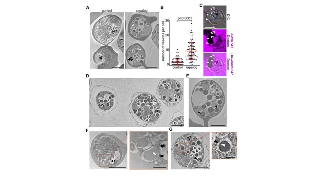
Vesicles Accumulating through Inactivation of VPS45 Are Filled with Host Cell Material (A) Representative transmission electron microscopy (EM) images of Percoll enriched PfVPS452xFKBP-FP parasites 8hr after induction of knock sideways (rapalog) compared with control. Arrowheads: examples of the induced vesicular structures. (B) Number of vesicles per cell section of EM images generated for(A) (n=101 cells for each, control and knock-sideways; error bars, SD; p-value is indicated, two-tailed, unpaired t-test). (C) Confocal microscopy images (single z-plane) of PfVPS45-2xFKBP-GFP parasites in red blood cells pre-loaded with AlexaFluor647 dextran. A Gauss filter was applied in Corel PHOTO-PAINTX6. Redline, outline of red blood cell; blue line, outline of parasite. Arrowheads, induced vesicles and their dextran content. FV, food vacuole; DIC, differential interference contrast. (D) EM images of PfVPS45-2xFKBP-GFP parasites 8hr after inducing the knock-sideways andt reatment with tetanolysin to release the host cell cytosol before embedding. (E) EM images of PfVPS45-2xFKBP-GFP parasites after knock-sideways, showing vesicles within the induced vesicles (arrowheads). (F and G) EM images showd ouble-membraned vesicles (F) after inactivation of PfVPS45 in parasites in intact red blood cells and (G) after release of host cell cytosol with tetanolysin. The orange frames show enlargements where white and black arrowheads indicate the inner and outer membrane of the vesicles, respectively; black arrowhead in the main image in (G) shows vesicle within a vesicle. Sizebars: (A), (D),and (E), 2mm; (C), 5mm; (F) and (G), 2 mm and enlargements 0.5mm.
Jonscher E, Flemming S, Schmitt M, Sabitzki R, Reichard N, Birnbaum J, Bergmann B, Höhn K, Spielmann T. PfVPS45 Is Required for Host Cell Cytosol Uptake by Malaria Blood Stage Parasites. Cell Host Microbe. 2019 25(1):166-173.e5.