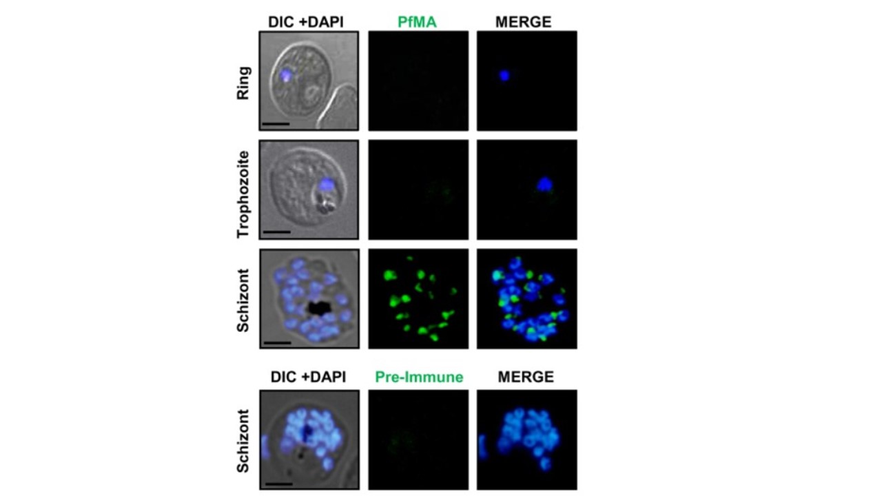
Protein expression of PfMA in different blood stages of P. falciparum. PfMA protein expression was analyzed in different stages of the asexual blood-stage life cycle by confocal immune-fluorescence microscopy using anti-PfMA mouse serum (green). Parasite nuclei were counterstained with DAPI (blue). No detectable PfMA staining was observed in ring and trophozoite stages. In schizonts, PfMA was detected as a punctate staining characteristic of many apical organelle resident proteins. No staining was detected with pre-immune sera. The scale bar indicates 2 μm. Hans N, Singh S, Pandey AK, Reddy KS, Gaur D, Chauhan VS. Identification and character-rization of a novel Plasmodium falciparum adhesin involved in erythrocyte invasion. PLoS One. 2013 8(9):e74790.
