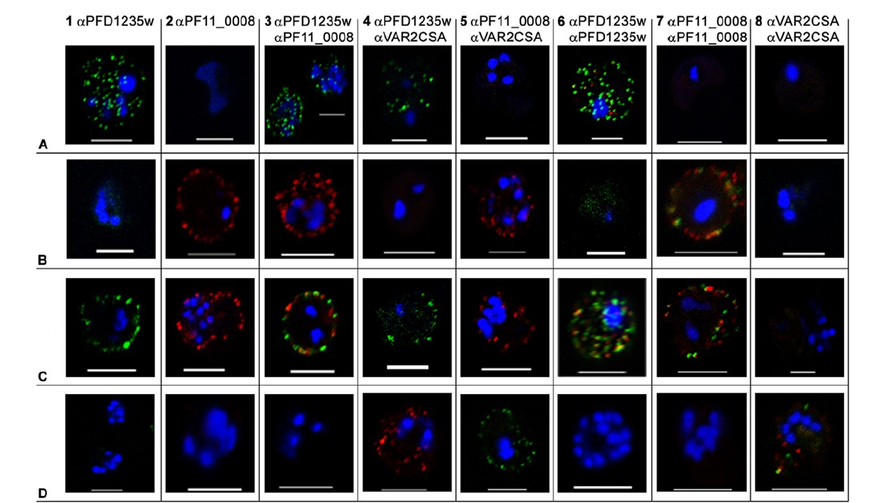
Surface expression of PfEMP1 on single 3D7 infected erythrocytes. (A) erythrocytes infected with a 3D7PFD1235w, (B) a 3D7PF11_0008, (C) a3D7PFD1235w/PF11_0008, and (D) a NF54 VAR2CSA sub-line. Localisation of PfEMP1 by confocal microscopy was done using (A1–D1) rat PFD1235w-DBL4c antisera and (A2–D2) rabbit PF11_0008-CIDR2b antisera. Double surface staining was done using the following combinations (A3–D3) rat PFD1235w-DBL4c and rabbit PF11_0008-CIDR2b antisera; (A4–D4) rat PFD1235w-DBL4c and mouse VAR2CSA-DBL5e antisera; (A5–D5) rabbit PF11_0008-CIDR2b and mouse VAR2CSA-DBL5e antisera; (A6–D6) rat PFD1235w-DBL4c and rabbit PFD1235w-CIDR1a; (A7–D7) rabbit PF11_0008-CIDR2b and rat PF11_0008-DBL4b; (A8–D8) rabbit VAR2CSA-DBL5e-DBL6e and mouse VAR2CSA-DBL5e antisera. Antisera staining of PFD1235w expressed by the 3D7PFD1235w (A1, A3, A4, A6) and 3D7PFD1235w/PF11_0008 (C1, C3, C4, C6) sub-lines was detected using a secondary antibody labelled with Alexa 488 (green). Antisera staining of PF11_0008 expressed by the 3D7PF11_0008 (B2, B3, B5, B7) and 3D7PFD1235w/PF11_0008 (C2, C3, C5, C7) was detected using a secondary antibody labelled with Alexa 568 (red). Staining of VAR2CSA expressed by NF54VAR2CSA is red (D4, D8) and green (D5, D8). DAPI staining of DNA in the nuclei is blue. Scale bar 5 mm.
Joergensen L, Bengtsson DC, Bengtsson A, Ronander E, Berger SS, Turner L, Dalgaard MB, Cham GK, Victor ME, Lavstsen T, Theander TG, Arnot DE, Jensen AT. Surface co-expression of two different PfEMP1 antigens on single plasmodium falciparum-infected erythrocytes facilitates binding to ICAM1 and PECAM1. PLoS Pathog. 2010 6(9):e1001083
Other associated proteins
| PFID | Formal Annotation |
|---|---|
| PF3D7_1100200 | erythrocyte membrane protein 1, PfEMP1 |
| PF3D7_1200600 | erythrocyte membrane protein 1, PfEMP1 |
