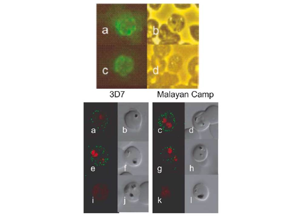
Localization of PIESP1 and PIESP2 with specific antibodies. (A) Immunofluorescent microscopy of PIEs (P. falciparum strain 3D7) fixed onto glass slides. Glass slide were incubated with anti-PIESP1 (Plates a and b), anti-PIESP2 (Plates c and d), and pooled preimmune sera (Plates e and f). Fluorescent images (Plates a, c, and e) and phase-contrast images (Plates b, d, and f) were collected simultaneously on an Olympus BX-60. (B) Localization of PIESPs with immunofluorescent confocal microscopy. Live P. falciparum strains 3D7 and Malayan Camp (unselected for knob and rosetting phenotypes) were incubated in suspension with specified antibodies. Plates a–d, anti-PIESP1 as primary antibody; Plates e–h, anti-PIESP2 as primary antibody; Plates i–l, preimmune sera as primary antibody. The 488 and 568 nm laser channels, for Alexa Fluor 488 and ethidine bromide, respectively, were collected separately and later superimposed (Plates a, c, e, g, i, and k). The DIC images (Plates b, d, f, h, j, and l) were collected simultaneously using the transmitted light detector.
Florens L, Liu X, Wang Y, Yang S, Schwartz O, Peglar M, Carucci DJ, Yates JR 3rd, Wub Y. Proteomics approach reveals novel proteins on the surface of malaria-infected erythrocytes. Mol Biochem Parasitol. 2004 135:1-11. Copyright Elsevier 2010.
Other associated proteins
| PFID | Formal Annotation |
|---|---|
| PF3D7_0310400 | TVN-junction protein 1 parasite-infected erythrocyte surface protein |
