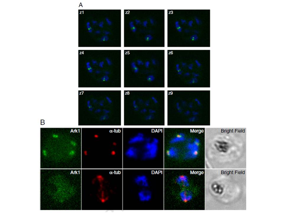
Pfark-1 expression in schizont stage P. falciparum parasites. A. Z-stacks live images of Pfark-1-GFP transgenic parasite undergoing schizogony showing the presence of pairs of dots (green) restricted to two out of seven nuclei (blue), reflecting the asynchrony of nuclear multiplication in schizont stage malaria parasites. B. Immunofluorescence assay of Pfark-1-GFP transgenic parasites. The upper panel shows that the Pfark-1-GFP protein is observed as pairs of dots (green) in close vicinity to DAPI-stained nuclear bodies (blue). Anti-α-tubulin antibody (DM1A) recognizes short intranuclear microtubule spindles (red) which appear between duplicated spindle pole bodies. The lower panel shows the absence of Pfark1-GFP protein at the spindle poles when fully developed spindle microtubules are visible.
Doerig C, Reininger L. Nima- and Aurora-related kinases of malaria parasites. Biochim Biophys Acta. 2013 1834(7):1336-45. Copyright Elsevier
Other associated proteins
| PFID | Formal Annotation |
|---|---|
| PF3D7_0903700 | alpha tubulin 1 |
