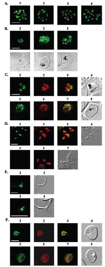
A, localization of RAMA in rhoptry organelles. Confocal microscopy on schizont stage parasites stained with rabbit anti-RAMA-B (1), anti-RAMA-C (2), anti-RAMA-D (3), or anti-RAMA-E (4) sera. B, localization of RAMA in immature parasites. Confocal microscopy on parasites stained with rabbit anti-RAMA-D serum: early trophozoite (1), late trophozoite (2), and early schizont (3) stages. Corresponding differential interference contrast (DIC) images are shown. C, trafficking of RAMA through the secretory pathway. Confocal microscopy on parasites stained with mouse anti-RAMA-E (1 and 5) and either anti-PfGRP (2) or anti-PfERD2 (6) rabbit antibodies. Corresponding overlay (3 and 7) and DIC (4 and 8) images are shown. D, localization of p60/RAMA in free merozoites. Confocal microscopy on parasites stained with mouse anti-MSP4 (2 and 6), and rabbit anti-RAMA-D (1) or anti-RAMA-B (5) sera. Corresponding overlay (3) and DIC (4 and 7) images are shown. E, discharge of RAMA from rhoptries. Confocal microscopy on CytB-treated merozoites incubated with RBCs, stained with rabbit anti-RAMA-D serum (1 and 3). Corresponding DIC images (2 and 4) are shown. F, association with the PV. Confocal microscopy on early ring stage-parasites stained with rabbit anti-RAMA-E serum (1 and 5) and monoclonal anti-RAP1 (2 and 6) antibodies. Corresponding overlay (3 and 7) and DIC (4 and 8) images are shown. Bars in 1 represent 5 mm. RAMA is synthesized as a 170-kDa protein in early trophozoites, several hours before rhoptry formation and is transiently localized within the endoplasmic reticulum and Golgi within lipid-rich microdomains.
Topolska AE, Lidgett A, Truman D, Fujioka H, Coppel RL. Characterization of a membrane-associated rhoptry protein of Plasmodium falciparum. J Biol Chem. 2004 279:4648-4656.
Other associated proteins
| PFID | Formal Annotation |
|---|---|
| PF3D7_0917900 | PfHsp70-2 |
| PF3D7_1353600 | ER lumen protein retaining receptor |
